| 登録情報 | データベース: PDB / ID: 7x4n
|
|---|
| タイトル | Crystal Structure of C. elegans kinesin-4 KLP-12 complexed with tubulin and DARPin |
|---|
 要素 要素 | - DARPin
- Kinesin-like protein
- Tubulin alpha-1A chain
- Tubulin beta chain
|
|---|
 キーワード キーワード | MOTOR PROTEIN / kinesin-4 / KLP-12 / KIF21A / KIF21B / microtubule / tubulin / axon / CFEOM1 / DARPin |
|---|
| 機能・相同性 |  機能・相同性情報 機能・相同性情報
COPI-dependent Golgi-to-ER retrograde traffic / Kinesins / kinesin complex / microtubule motor activity / motile cilium / microtubule-based movement / structural constituent of cytoskeleton / 加水分解酵素; 酸無水物に作用; 酸無水物に作用・細胞または細胞小器官の運動に関与 / microtubule cytoskeleton organization / neuron migration ...COPI-dependent Golgi-to-ER retrograde traffic / Kinesins / kinesin complex / microtubule motor activity / motile cilium / microtubule-based movement / structural constituent of cytoskeleton / 加水分解酵素; 酸無水物に作用; 酸無水物に作用・細胞または細胞小器官の運動に関与 / microtubule cytoskeleton organization / neuron migration / mitotic cell cycle / microtubule binding / 加水分解酵素; 酸無水物に作用; GTPに作用・細胞または細胞小器官の運動に関与 / microtubule / hydrolase activity / GTPase activity / GTP binding / ATP hydrolysis activity / ATP binding / metal ion binding / cytoplasm類似検索 - 分子機能 Kinesin motor domain / Kinesin / Kinesin-like protein / Tubulin/FtsZ, C-terminal domain / Tubulin/FtsZ, GTPase domain / Kinesin motor domain signature. / Kinesin motor domain, conserved site / Kinesin motor domain / Kinesin motor domain profile. / Kinesin motor, catalytic domain. ATPase. ...Kinesin motor domain / Kinesin / Kinesin-like protein / Tubulin/FtsZ, C-terminal domain / Tubulin/FtsZ, GTPase domain / Kinesin motor domain signature. / Kinesin motor domain, conserved site / Kinesin motor domain / Kinesin motor domain profile. / Kinesin motor, catalytic domain. ATPase. / Kinesin motor domain / 60s Ribosomal Protein L30; Chain: A; / Kinesin motor domain superfamily / Tubulin-beta mRNA autoregulation signal. / Alpha tubulin / Beta tubulin, autoregulation binding site / Beta tubulin / Tubulin / Tubulin, C-terminal / Tubulin C-terminal domain / Tubulin, conserved site / Tubulin subunits alpha, beta, and gamma signature. / Tubulin/FtsZ family, C-terminal domain / Tubulin/FtsZ-like, C-terminal domain / Tubulin/FtsZ, C-terminal / Tubulin/FtsZ, 2-layer sandwich domain / Tubulin/FtsZ family, GTPase domain / Tubulin/FtsZ family, GTPase domain / Tubulin/FtsZ, GTPase domain / Tubulin/FtsZ, GTPase domain superfamily / WD domain, G-beta repeat / WD40 repeat, conserved site / Trp-Asp (WD) repeats signature. / Trp-Asp (WD) repeats profile. / Trp-Asp (WD) repeats circular profile. / WD40 repeats / WD40 repeat / WD40-repeat-containing domain superfamily / WD40/YVTN repeat-like-containing domain superfamily / Rossmann fold / P-loop containing nucleoside triphosphate hydrolase / 2-Layer Sandwich / 3-Layer(aba) Sandwich / Alpha Beta類似検索 - ドメイン・相同性 PHOSPHOAMINOPHOSPHONIC ACID-ADENYLATE ESTER / GUANOSINE-5'-DIPHOSPHATE / GUANOSINE-5'-TRIPHOSPHATE / Kinesin-like protein klp-12 / Tubulin alpha-1A chain / Tubulin beta chain類似検索 - 構成要素 |
|---|
| 生物種 | synthetic construct (人工物)
  Caenorhabditis elegans (センチュウ) Caenorhabditis elegans (センチュウ)
  Sus scrofa (ブタ) Sus scrofa (ブタ) |
|---|
| 手法 |  X線回折 / シンクロトロン / 分子置換 / 解像度: 2.88 Å X線回折 / シンクロトロン / 分子置換 / 解像度: 2.88 Å |
|---|
 データ登録者 データ登録者 | Taguchi, S. / Imasaki, T. / Saijo-Hamano, Y. / Sakai, N. / Nitta, R. |
|---|
| 資金援助 |  日本, 5件 日本, 5件 | 組織 | 認可番号 | 国 |
|---|
| Japan Agency for Medical Research and Development (AMED) | JP21am0101070 | 日本 | | Japan Society for the Promotion of Science (JSPS) | 19K07246 | 日本 | | Japan Society for the Promotion of Science (JSPS) | 19H03396 | 日本 | | Japan Agency for Medical Research and Development (AMED) | JP21gm0810013 | 日本 | | Japan Science and Technology | JPMJMS2024 | 日本 |
|
|---|
 引用 引用 | ジャーナル: Elife / 年: 2022
タイトル: Structural model of microtubule dynamics inhibition by kinesin-4 from the crystal structure of KLP-12 -tubulin complex.
著者: Taguchi, S. / Nakano, J. / Imasaki, T. / Kita, T. / Saijo-Hamano, Y. / Sakai, N. / Shigematsu, H. / Okuma, H. / Shimizu, T. / Nitta, E. / Kikkawa, S. / Mizobuchi, S. / Niwa, S. / Nitta, R. |
|---|
| 履歴 | | 登録 | 2022年3月3日 | 登録サイト: PDBJ / 処理サイト: PDBJ |
|---|
| 改定 1.0 | 2022年9月21日 | Provider: repository / タイプ: Initial release |
|---|
| 改定 1.1 | 2022年9月28日 | Group: Database references / カテゴリ: citation / citation_author / Item: _citation.pdbx_database_id_PubMed / _citation.title |
|---|
| 改定 1.2 | 2023年11月29日 | Group: Data collection / Refinement description
カテゴリ: chem_comp_atom / chem_comp_bond / pdbx_initial_refinement_model |
|---|
|
|---|
 ムービー
ムービー コントローラー


 データを開く
 基本情報
基本情報 構造の表示
構造の表示 ダウンロードとリンク
 その他のダウンロード
 PDBj
PDBj


 集合体
集合体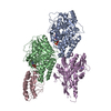


 分子量: 523.180 Da / 分子数: 1 / 由来タイプ: 合成 / 式: C10H16N5O14P3 / タイプ: SUBJECT OF INVESTIGATION / コメント: GTP, エネルギー貯蔵分子*YM
 分子量: 24.305 Da / 分子数: 2 / 由来タイプ: 合成 / 式: Mg / タイプ: SUBJECT OF INVESTIGATION
 タイプ: RNA linking / 分子量: 443.201 Da / 分子数: 1 / 由来タイプ: 合成 / 式: C10H15N5O11P2 / タイプ: SUBJECT OF INVESTIGATION / コメント: GDP, エネルギー貯蔵分子*YM
 分子量: 506.196 Da / 分子数: 1 / 由来タイプ: 合成 / 式: C10H17N6O12P3 / タイプ: SUBJECT OF INVESTIGATION
 試料調製
試料調製 解析
解析