 Movie
Movie Controller


+ Open data

- Basic information
| Entry | Database: PDB / ID: 7x3n | |||||||||
|---|---|---|---|---|---|---|---|---|---|---|
| Title | Crystal structure of anti-mPEG h15-2b Fab | |||||||||
 Components Components |
| |||||||||
 Keywords Keywords | IMMUNE SYSTEM / methoxy polyethylene glycol / mPEG / anti-mPEG antibody | |||||||||
| Function / homology | Immunoglobulins / Immunoglobulin-like / Sandwich / Mainly Beta / 2,5,8,11,14,17-HEXAOXANONADECAN-19-OL Function and homology information Function and homology information | |||||||||
| Biological species |  Homo sapiens (human) Homo sapiens (human) | |||||||||
| Method |  X-RAY DIFFRACTION / SYNCHROTRON / MOLECULAR REPLACEMENT / Resolution: 2.24 Å X-RAY DIFFRACTION / SYNCHROTRON / MOLECULAR REPLACEMENT / Resolution: 2.24 Å | |||||||||
 Authors Authors | Chang, C.Y. / Nguyen, T.M.T. / Toh, S.I. / Su, Y.C. | |||||||||
| Funding support |  Taiwan, 2items Taiwan, 2items
| |||||||||
 Citation Citation | Journal: Commun Chem / Year: 2022 Title: Structural determination of an antibody that specifically recognizes polyethylene glycol with a terminal methoxy group Authors: Nguyen, M.T.T. / Shih, Y.C. / Lin, M.H. / Roffler, S.R. / Hsiao, C.Y. / Cheng, T.L. / Lin, W.W. / Lin, E.C. / Jong, Y.J. / Chang, C.Y. / Su, Y.C. | |||||||||
| History |
|
- Structure visualization
| Structure viewer | Molecule: MolmilJmol/JSmol |
|---|
- Downloads & links
-Download
| PDBx/mmCIF format | 7x3n.cif.gz | 179.7 KB | Display | PDBx/mmCIF format |
|---|---|---|---|---|
| PDB format | pdb7x3n.ent.gz | 140.7 KB | Display | PDB format |
| PDBx/mmJSON format | 7x3n.json.gz | Tree view | PDBx/mmJSON format | |
| Others |  Other downloads Other downloads |
-Validation report
| Arichive directory | https://data.pdbj.org/pub/pdb/validation_reports/x3/7x3nftp://data.pdbj.org/pub/pdb/validation_reports/x3/7x3n | HTTPS FTP |
|---|
-Related structure data
| Related structure data |  1l7iS S: Starting model for refinement |
|---|---|
| Similar structure data |
-Links
 PDBj
PDBj

- Assembly
| Deposited unit | 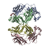
| ||||||||
|---|---|---|---|---|---|---|---|---|---|
| 1 | 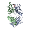
| ||||||||
| 2 | 
| ||||||||
| Unit cell |
|
-Components
| #1: Antibody | Mass: 23539.170 Da / Num. of mol.: 2 Source method: isolated from a genetically manipulated source Source: (gene. exp.) Homo sapiens (human) / Production host:   Cricetulus griseus (Chinese hamster) Cricetulus griseus (Chinese hamster)#2: Antibody | Mass: 24598.355 Da / Num. of mol.: 2 Source method: isolated from a genetically manipulated source Source: (gene. exp.) Homo sapiens (human) / Production host: Cricetulus griseus (Chinese hamster)#3: Chemical |   Mass: 296.357 Da / Num. of mol.: 2 / Source method: obtained synthetically / Formula: C13H28O7 / Feature type: SUBJECT OF INVESTIGATION Mass: 296.357 Da / Num. of mol.: 2 / Source method: obtained synthetically / Formula: C13H28O7 / Feature type: SUBJECT OF INVESTIGATION#4: Water | ChemComp-HOH / |  Mass: 18.015 Da / Num. of mol.: 346 / Source method: isolated from a natural source / Formula: H2O Mass: 18.015 Da / Num. of mol.: 346 / Source method: isolated from a natural source / Formula: H2OHas ligand of interest | Y | Has protein modification | Y | |
|---|
-Experimental details
-Experiment
| Experiment | Method: X-RAY DIFFRACTION / Number of used crystals: 1 |
|---|
- Sample preparation
| Crystal | Density Matthews: 2.97 Å3/Da / Density % sol: 58.56 % |
|---|---|
| Crystal grow | Temperature: 291 K / Method: vapor diffusion, hanging drop Details: 18% (w/v) PEG 6000, 1% (w/v) PEG 2000 MME, 0.15 M lithium sulfate monohydrate, 0.1 M citric acid, pH 3.5 |
-Data collection
| Diffraction | Mean temperature: 80 K / Serial crystal experiment: N |
|---|---|
| Diffraction source | Source: SYNCHROTRON / Site: NSRRC / Beamline: BL13B1 / Wavelength: 1 Å |
| Detector | Type: ADSC QUANTUM 315r / Detector: CCD / Date: Jun 10, 2020 |
| Radiation | Protocol: SINGLE WAVELENGTH / Monochromatic (M) / Laue (L): M / Scattering type: x-ray |
| Radiation wavelength | Wavelength: 1 Å / Relative weight: 1 |
| Reflection | Resolution: 2.24→30 Å / Num. obs: 55882 / % possible obs: 99.7 % / Redundancy: 7.1 % / CC1/2: 0.926 / Net I/σ(I): 12.7 |
| Reflection shell | Resolution: 2.24→2.34 Å / Num. unique obs: 6846 / CC1/2: 0.816 / % possible all: 99.6 |
- Processing
| Software |
| ||||||||||||||||
|---|---|---|---|---|---|---|---|---|---|---|---|---|---|---|---|---|---|
| Refinement | Method to determine structure: MOLECULAR REPLACEMENT Starting model: 1L7I Resolution: 2.24→27.61 Å / Cross valid method: THROUGHOUT
| ||||||||||||||||
| Displacement parameters | Biso max: 88.75 Å2 / Biso mean: 25.7941 Å2 / Biso min: 4.11 Å2 | ||||||||||||||||
| Refinement step | Cycle: LAST / Resolution: 2.24→27.61 Å
| ||||||||||||||||
| LS refinement shell | Resolution: 2.24→2.34 Å /
|
