 Movie
Movie Controller


+ Open data

- Basic information
| Entry | Database: PDB / ID: 1l7i | ||||||
|---|---|---|---|---|---|---|---|
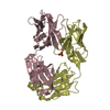
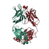
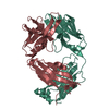
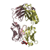
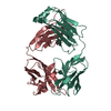
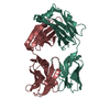
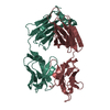
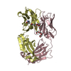
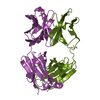
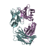
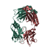
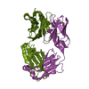
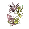
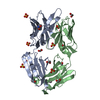
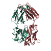
| Title | Crystal Structure of the anti-ErbB2 Fab2C4 | ||||||
 Components Components | (chimera of Fab2C4: "humanized" murine monoclonal antibody) x 2 | ||||||
 Keywords Keywords | IMMUNE SYSTEM / Ig domain / Fab fragment | ||||||
| Function / homology |  Function and homology information Function and homology informationimmunoglobulin complex / adaptive immune response / extracellular region / plasma membrane Similarity search - Function | ||||||
| Biological species |  Homo sapiens (human) Homo sapiens (human) | ||||||
| Method |  X-RAY DIFFRACTION / SYNCHROTRON / MOLECULAR REPLACEMENT / Resolution: 1.8 Å X-RAY DIFFRACTION / SYNCHROTRON / MOLECULAR REPLACEMENT / Resolution: 1.8 Å | ||||||
 Authors Authors | Vajdos, F.F. / Adams, C.W. / Breece, T.N. / Presta, L.G. / de Vos, A.M. / Sidhu, S.S. | ||||||
 Citation Citation | Journal: J.Mol.Biol. / Year: 2002 Title: Comprehensive functional maps of the antigen-binding site of an anti-ErbB2 antibody obtained with shotgun scanning mutagenesis. Authors: Vajdos, F.F. / Adams, C.W. / Breece, T.N. / Presta, L.G. / de Vos, A.M. / Sidhu, S.S. | ||||||
| History |
| ||||||
| Remark 999 | sequence an appropriate sequence database reference was not available at the time of processing. ...sequence an appropriate sequence database reference was not available at the time of processing. Fab2C4 is a "humanized" murine monoclonal antibody, with the following residues corresponding to the human Ig sequence: L1-L23, L35-L49, L57-L88, L98-L214, H1-H25, H36-H49, H66-H68, H70, H72, H74-H94, H102-223. RESIDUES L24-L34, L50-L56, L89-L97, H26-H35, H50-H65, H69, H71, H73, H95-H102 CORRESPOND TO THE ORIGINAL MURINE SEQUENCE. |
- Structure visualization
| Structure viewer | Molecule: MolmilJmol/JSmol |
|---|
- Downloads & links
-Download
| PDBx/mmCIF format | 1l7i.cif.gz | 105.4 KB | Display | PDBx/mmCIF format |
|---|---|---|---|---|
| PDB format | pdb1l7i.ent.gz | 79.1 KB | Display | PDB format |
| PDBx/mmJSON format | 1l7i.json.gz | Tree view | PDBx/mmJSON format | |
| Others |  Other downloads Other downloads |
-Validation report
| Arichive directory | https://data.pdbj.org/pub/pdb/validation_reports/l7/1l7iftp://data.pdbj.org/pub/pdb/validation_reports/l7/1l7i | HTTPS FTP |
|---|
-Related structure data
| Similar structure data |
|---|


-Links
 PDBj
PDBj

- Assembly
| Deposited unit | 
| ||||||||
|---|---|---|---|---|---|---|---|---|---|
| 1 |
| ||||||||
| Unit cell |
| ||||||||
| Details | The biological assembly is a heterodimer of heavy and light chains. |
-Components
| #1: Antibody | Mass: 23548.152 Da / Num. of mol.: 1 / Fragment: light chain (residues 1-214) Source method: isolated from a genetically manipulated source Details: The chimera consists of the human Ig and the original murine sequence Source: (gene. exp.) Homo sapiens (human) / Production host:  | ||||
|---|---|---|---|---|---|
| #2: Antibody | Mass: 23674.486 Da / Num. of mol.: 1 / Fragment: heavy chain (residues 1-216) Source method: isolated from a genetically manipulated source Details: The chimera consists of the human Ig and the original murine sequence Source: (gene. exp.) Homo sapiens (human) / Production host: | ||||
| #3: Chemical |   Mass: 96.063 Da / Num. of mol.: 2 / Source method: obtained synthetically / Formula: SO4 Mass: 96.063 Da / Num. of mol.: 2 / Source method: obtained synthetically / Formula: SO4#4: Water | ChemComp-HOH / |  Mass: 18.015 Da / Num. of mol.: 392 / Source method: isolated from a natural source / Formula: H2O Mass: 18.015 Da / Num. of mol.: 392 / Source method: isolated from a natural source / Formula: H2OHas protein modification | Y | |
-Experimental details
-Experiment
| Experiment | Method: X-RAY DIFFRACTION / Number of used crystals: 1 |
|---|
- Sample preparation
| Crystal | Density Matthews: 2.19 Å3/Da / Density % sol: 43.71 % | ||||||||||||||||||||||||
|---|---|---|---|---|---|---|---|---|---|---|---|---|---|---|---|---|---|---|---|---|---|---|---|---|---|
| Crystal grow | Temperature: 292 K / Method: vapor diffusion, hanging drop / pH: 8 Details: 35%-45% saturated (NH4)2SO4, 0.1 M Tris-HCl, pH 8.0, VAPOR DIFFUSION, HANGING DROP, temperature 292K | ||||||||||||||||||||||||
| Crystal grow | *PLUS Method: vapor diffusion, sitting drop / Details: used microseeding | ||||||||||||||||||||||||
| Components of the solutions | *PLUS
|
-Data collection
| Diffraction | Mean temperature: 100 K |
|---|---|
| Diffraction source | Source: SYNCHROTRON / Site: ALS  / Beamline: 5.0.2 / Wavelength: 1.1 Å / Beamline: 5.0.2 / Wavelength: 1.1 Å |
| Detector | Type: ADSC QUANTUM 4 / Detector: CCD / Date: Oct 30, 2000 |
| Radiation | Monochromator: Double crystal / Protocol: SINGLE WAVELENGTH / Monochromatic (M) / Laue (L): M / Scattering type: x-ray |
| Radiation wavelength | Wavelength: 1.1 Å / Relative weight: 1 |
| Reflection | Resolution: 1.8→15 Å / Num. all: 36884 / Num. obs: 36884 / % possible obs: 97.6 % / Observed criterion σ(F): 0 / Observed criterion σ(I): 0 / Redundancy: 2.3 % / Biso Wilson estimate: 14.9 Å2 / Rmerge(I) obs: 0.065 / Net I/σ(I): 5.2 |
| Reflection shell | Resolution: 1.8→1.9 Å / Rmerge(I) obs: 0.328 / Mean I/σ(I) obs: 1.4 / % possible all: 97.6 |
| Reflection | *PLUS Lowest resolution: 15 Å / Num. measured all: 85734 |
| Reflection shell | *PLUS % possible obs: 97.6 % / Redundancy: 2.3 % |
- Processing
| Software |
| ||||||||||||||||||||||||||||||||||||
|---|---|---|---|---|---|---|---|---|---|---|---|---|---|---|---|---|---|---|---|---|---|---|---|---|---|---|---|---|---|---|---|---|---|---|---|---|---|
| Refinement | Method to determine structure: MOLECULAR REPLACEMENT / Resolution: 1.8→14.39 Å / Rfactor Rfree error: 0.005 / Data cutoff high absF: 1429863.38 / Data cutoff low absF: 0 / Isotropic thermal model: RESTRAINED / Cross valid method: THROUGHOUT / σ(F): 0 / Stereochemistry target values: Engh & Huber
| ||||||||||||||||||||||||||||||||||||
| Displacement parameters | Biso mean: 24 Å2
| ||||||||||||||||||||||||||||||||||||
| Refine analyze |
| ||||||||||||||||||||||||||||||||||||
| Refinement step | Cycle: LAST / Resolution: 1.8→14.39 Å
| ||||||||||||||||||||||||||||||||||||
| Refine LS restraints |
| ||||||||||||||||||||||||||||||||||||
| LS refinement shell | Resolution: 1.8→1.91 Å / Rfactor Rfree error: 0.021 / Total num. of bins used: 6
| ||||||||||||||||||||||||||||||||||||
| Xplor file |
| ||||||||||||||||||||||||||||||||||||
| Refinement | *PLUS % reflection Rfree: 5 % / Rfactor Rfree: 0.23 | ||||||||||||||||||||||||||||||||||||
| Solvent computation | *PLUS | ||||||||||||||||||||||||||||||||||||
| Displacement parameters | *PLUS | ||||||||||||||||||||||||||||||||||||
| Refine LS restraints | *PLUS
| ||||||||||||||||||||||||||||||||||||
| LS refinement shell | *PLUS Rfactor Rwork: 0.27 |
