 Movie
Movie Controller

[English] 日本語
 Yorodumi
- PDB-7x17: Structure of Pseudomonas NRPS protein, AmbB-TC bound to Ppant-L-Ala -
+ Open data

- Basic information
| Entry | Database: PDB / ID: 7x17 | ||||||
|---|---|---|---|---|---|---|---|
| Title | Structure of Pseudomonas NRPS protein, AmbB-TC bound to Ppant-L-Ala | ||||||
 Components Components | AMB antimetabolite synthase AmbB | ||||||
 Keywords Keywords | BIOSYNTHETIC PROTEIN / Non-ribosomal peptide synthetase / AmbB / Pseudomonas | ||||||
| Function / homology |  Function and homology information Function and homology informationL-alanine-[L-alanyl-carrier protein] ligase / 2,3-dihydroxybenzoate-serine ligase activity / enterobactin synthetase complex / enterobactin biosynthetic process / amino acid activation for nonribosomal peptide biosynthetic process / phosphopantetheine binding / cytoplasm / cytosol Similarity search - Function | ||||||
| Biological species |  Pseudomonas aeruginosa PAO1 (bacteria) Pseudomonas aeruginosa PAO1 (bacteria) | ||||||
| Method |  X-RAY DIFFRACTION / SYNCHROTRON / MOLECULAR REPLACEMENT / Resolution: 2.5 Å X-RAY DIFFRACTION / SYNCHROTRON / MOLECULAR REPLACEMENT / Resolution: 2.5 Å | ||||||
 Authors Authors | ChuYuanKee, M. / Bharath, S.R. / Song, H. | ||||||
| Funding support | 1items
| ||||||
 Citation Citation | Journal: Sci Rep / Year: 2022 Title: Structural insights into the substrate-bound condensation domains of non-ribosomal peptide synthetase AmbB. Authors: Chu Yuan Kee, M.J. / Bharath, S.R. / Wee, S. / Bowler, M.W. / Gunaratne, J. / Pan, S. / Zhang, L. / Song, H. | ||||||
| History |
|
- Structure visualization
| Structure viewer | Molecule: MolmilJmol/JSmol |
|---|
- Downloads & links
-Download
| PDBx/mmCIF format | 7x17.cif.gz | 426.4 KB | Display | PDBx/mmCIF format |
|---|---|---|---|---|
| PDB format | pdb7x17.ent.gz | 278 KB | Display | PDB format |
| PDBx/mmJSON format | 7x17.json.gz | Tree view | PDBx/mmJSON format | |
| Others |  Other downloads Other downloads |
-Validation report
| Arichive directory | https://data.pdbj.org/pub/pdb/validation_reports/x1/7x17ftp://data.pdbj.org/pub/pdb/validation_reports/x1/7x17 | HTTPS FTP |
|---|
-Related structure data
| Related structure data | 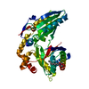 7x0eSC 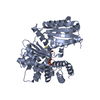 7x0fC S: Starting model for refinement C: citing same article ( |
|---|---|
| Similar structure data |
-Links
 PDBj
PDBj


- Assembly
| Deposited unit | 
| ||||||||||
|---|---|---|---|---|---|---|---|---|---|---|---|
| 1 | 
| ||||||||||
| 2 | 
| ||||||||||
| 3 | 
| ||||||||||
| 4 | 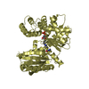
| ||||||||||
| Unit cell |
|
-Components
| #1: Protein | Mass: 55970.652 Da / Num. of mol.: 4 Source method: isolated from a genetically manipulated source Source: (gene. exp.) Pseudomonas aeruginosa PAO1 (bacteria) / Gene: ambB, PA2305 / Production host: References: UniProt: Q9I1H0, L-alanine-[L-alanyl-carrier protein] ligase #2: Chemical | ChemComp-868 / 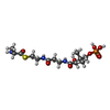  Mass: 429.426 Da / Num. of mol.: 4 / Source method: obtained synthetically / Formula: C14H28N3O8PS / Feature type: SUBJECT OF INVESTIGATION Mass: 429.426 Da / Num. of mol.: 4 / Source method: obtained synthetically / Formula: C14H28N3O8PS / Feature type: SUBJECT OF INVESTIGATION#3: Water | ChemComp-HOH / |  Mass: 18.015 Da / Num. of mol.: 416 / Source method: isolated from a natural source / Formula: H2O Mass: 18.015 Da / Num. of mol.: 416 / Source method: isolated from a natural source / Formula: H2OHas ligand of interest | Y | Has protein modification | Y | |
|---|
-Experimental details
-Experiment
| Experiment | Method: X-RAY DIFFRACTION / Number of used crystals: 1 |
|---|
- Sample preparation
| Crystal | Density Matthews: 2.59 Å3/Da / Density % sol: 52.5 % |
|---|---|
| Crystal grow | Temperature: 293 K / Method: vapor diffusion Details: 0.1M MES pH 6.6, 0.2M ammonium nitrate, 15% PEG 3350, 5% glycerol |
-Data collection
| Diffraction | Mean temperature: 100 K / Serial crystal experiment: N |
|---|---|
| Diffraction source | Source: SYNCHROTRON / Site: NSRRC  / Beamline: TPS 05A / Wavelength: 1 Å / Beamline: TPS 05A / Wavelength: 1 Å |
| Detector | Type: MAR scanner 300 mm plate / Detector: IMAGE PLATE / Date: Mar 15, 2019 |
| Radiation | Protocol: SINGLE WAVELENGTH / Monochromatic (M) / Laue (L): M / Scattering type: x-ray |
| Radiation wavelength | Wavelength: 1 Å / Relative weight: 1 |
| Reflection | Resolution: 2.5→48.83 Å / Num. obs: 78940 / % possible obs: 99.2 % / Redundancy: 3.4 % / Biso Wilson estimate: 24.52 Å2 / CC1/2: 0.997 / Net I/σ(I): 11.4 |
| Reflection shell | Resolution: 2.5→2.55 Å / Mean I/σ(I) obs: 1.7 / Num. unique obs: 4499 / CC1/2: 0.559 |
- Processing
| Software |
| |||||||||||||||||||||||||||||||||||||||||||||||||||||||||||||||||||||||||||||||||||||||||||||||||||||||||||||||||||||||||||||||||||||||||||||||||||||||||||||||||||||||||||||||||||||||||||||||||||||||||||
|---|---|---|---|---|---|---|---|---|---|---|---|---|---|---|---|---|---|---|---|---|---|---|---|---|---|---|---|---|---|---|---|---|---|---|---|---|---|---|---|---|---|---|---|---|---|---|---|---|---|---|---|---|---|---|---|---|---|---|---|---|---|---|---|---|---|---|---|---|---|---|---|---|---|---|---|---|---|---|---|---|---|---|---|---|---|---|---|---|---|---|---|---|---|---|---|---|---|---|---|---|---|---|---|---|---|---|---|---|---|---|---|---|---|---|---|---|---|---|---|---|---|---|---|---|---|---|---|---|---|---|---|---|---|---|---|---|---|---|---|---|---|---|---|---|---|---|---|---|---|---|---|---|---|---|---|---|---|---|---|---|---|---|---|---|---|---|---|---|---|---|---|---|---|---|---|---|---|---|---|---|---|---|---|---|---|---|---|---|---|---|---|---|---|---|---|---|---|---|---|---|---|---|---|---|
| Refinement | Method to determine structure: MOLECULAR REPLACEMENT Starting model: 7X0E Resolution: 2.5→46.59 Å / SU ML: 0.3807 / Cross valid method: FREE R-VALUE / σ(F): 1.36 / Phase error: 30.8858 Stereochemistry target values: GeoStd + Monomer Library + CDL v1.2
| |||||||||||||||||||||||||||||||||||||||||||||||||||||||||||||||||||||||||||||||||||||||||||||||||||||||||||||||||||||||||||||||||||||||||||||||||||||||||||||||||||||||||||||||||||||||||||||||||||||||||||
| Solvent computation | Shrinkage radii: 0.9 Å / VDW probe radii: 1.1 Å / Solvent model: FLAT BULK SOLVENT MODEL | |||||||||||||||||||||||||||||||||||||||||||||||||||||||||||||||||||||||||||||||||||||||||||||||||||||||||||||||||||||||||||||||||||||||||||||||||||||||||||||||||||||||||||||||||||||||||||||||||||||||||||
| Displacement parameters | Biso mean: 29.8 Å2 | |||||||||||||||||||||||||||||||||||||||||||||||||||||||||||||||||||||||||||||||||||||||||||||||||||||||||||||||||||||||||||||||||||||||||||||||||||||||||||||||||||||||||||||||||||||||||||||||||||||||||||
| Refinement step | Cycle: LAST / Resolution: 2.5→46.59 Å
| |||||||||||||||||||||||||||||||||||||||||||||||||||||||||||||||||||||||||||||||||||||||||||||||||||||||||||||||||||||||||||||||||||||||||||||||||||||||||||||||||||||||||||||||||||||||||||||||||||||||||||
| Refine LS restraints |
| |||||||||||||||||||||||||||||||||||||||||||||||||||||||||||||||||||||||||||||||||||||||||||||||||||||||||||||||||||||||||||||||||||||||||||||||||||||||||||||||||||||||||||||||||||||||||||||||||||||||||||
| LS refinement shell |
|
