| 登録情報 | データベース: PDB / ID: 7wz6
|
|---|
| タイトル | Crystal structure of MyoD-E47 |
|---|
 要素 要素 | - Isoform E47 of Transcription factor E2-alpha
- Myoblast determination protein 1
|
|---|
 キーワード キーワード | TRANSCRIPTION / E-box / bHLH domain |
|---|
| 機能・相同性 |  機能・相同性情報 機能・相同性情報
myoblast fate determination / skeletal muscle fiber adaptation / negative regulation of myoblast proliferation / myotube differentiation involved in skeletal muscle regeneration / positive regulation of snRNA transcription by RNA polymerase II / positive regulation of skeletal muscle tissue regeneration / myotube differentiation / bHLH transcription factor binding / cell development / RUNX1 regulates transcription of genes involved in differentiation of HSCs ...myoblast fate determination / skeletal muscle fiber adaptation / negative regulation of myoblast proliferation / myotube differentiation involved in skeletal muscle regeneration / positive regulation of snRNA transcription by RNA polymerase II / positive regulation of skeletal muscle tissue regeneration / myotube differentiation / bHLH transcription factor binding / cell development / RUNX1 regulates transcription of genes involved in differentiation of HSCs / lymphocyte differentiation / natural killer cell differentiation / immunoglobulin V(D)J recombination / Peyer's patch development / Myogenesis / cardiac muscle cell differentiation / skeletal muscle tissue regeneration / cellular response to oxygen levels / myoblast fusion / muscle cell differentiation / myoblast differentiation / cellular response to glucocorticoid stimulus / positive regulation of myoblast fusion / muscle organ development / positive regulation of muscle cell differentiation / DNA-binding transcription activator activity / B cell lineage commitment / regulation of alternative mRNA splicing, via spliceosome / myofibril / E-box binding / regulation of G1/S transition of mitotic cell cycle / skeletal muscle cell differentiation / positive regulation of DNA-binding transcription factor activity / skeletal muscle tissue development / cis-regulatory region sequence-specific DNA binding / skeletal muscle fiber development / positive regulation of cell cycle / gastrulation / positive regulation of B cell proliferation / striated muscle cell differentiation / positive regulation of neuron differentiation / cellular response to starvation / nuclear receptor binding / erythrocyte differentiation / promoter-specific chromatin binding / cellular response to estradiol stimulus / euchromatin / chromatin DNA binding / RNA polymerase II transcription regulator complex / cellular response to tumor necrosis factor / nucleosome / nervous system development / T cell differentiation in thymus / regulation of gene expression / DNA-binding transcription activator activity, RNA polymerase II-specific / response to lipopolysaccharide / gene expression / transcription regulator complex / DNA-binding transcription factor binding / sequence-specific DNA binding / RNA polymerase II-specific DNA-binding transcription factor binding / transcription by RNA polymerase II / DNA-binding transcription factor activity, RNA polymerase II-specific / protein stabilization / RNA polymerase II cis-regulatory region sequence-specific DNA binding / chromatin remodeling / response to xenobiotic stimulus / DNA-binding transcription factor activity / protein heterodimerization activity / ubiquitin protein ligase binding / chromatin binding / positive regulation of gene expression / regulation of DNA-templated transcription / regulation of transcription by RNA polymerase II / chromatin / positive regulation of DNA-templated transcription / enzyme binding / protein homodimerization activity / positive regulation of transcription by RNA polymerase II / protein-containing complex / DNA binding / nucleoplasm / identical protein binding / nucleus / cytoplasm / cytosol類似検索 - 分子機能 Myogenic muscle-specific protein, N-terminal / Myogenic determination factor 5 / Myogenic factor / Myogenic Basic domain / Myogenic determination factor 5 / Basic domain in HLH proteins of MYOD family / : / Helix-loop-helix DNA-binding domain / helix loop helix domain / Myc-type, basic helix-loop-helix (bHLH) domain ...Myogenic muscle-specific protein, N-terminal / Myogenic determination factor 5 / Myogenic factor / Myogenic Basic domain / Myogenic determination factor 5 / Basic domain in HLH proteins of MYOD family / : / Helix-loop-helix DNA-binding domain / helix loop helix domain / Myc-type, basic helix-loop-helix (bHLH) domain / Myc-type, basic helix-loop-helix (bHLH) domain profile. / Helix-loop-helix DNA-binding domain superfamily類似検索 - ドメイン・相同性 Myoblast determination protein 1 / Transcription factor E2-alpha類似検索 - 構成要素 |
|---|
| 生物種 |   Mus musculus (ハツカネズミ) Mus musculus (ハツカネズミ) |
|---|
| 手法 |  X線回折 / シンクロトロン / 分子置換 / 解像度: 2.05 Å X線回折 / シンクロトロン / 分子置換 / 解像度: 2.05 Å |
|---|
 データ登録者 データ登録者 | Zhong, J. / Huang, Y. / Ma, J. |
|---|
| 資金援助 |  中国, 1件 中国, 1件 | 組織 | 認可番号 | 国 |
|---|
| National Natural Science Foundation of China (NSFC) | 32171186 | 中国 |
|
|---|
 引用 引用 | ジャーナル: Biochem.Biophys.Res.Commun. / 年: 2022
タイトル: Structural basis of the bHLH domains of MyoD-E47 heterodimer.
著者: Zhong, J. / Jin, Z. / Jiang, L. / Zhang, L. / Hu, Z. / Zhang, Y. / Liu, Y. / Ma, J. / Huang, Y. |
|---|
| 履歴 | | 登録 | 2022年2月17日 | 登録サイト: PDBJ / 処理サイト: PDBJ |
|---|
| 改定 1.0 | 2022年6月22日 | Provider: repository / タイプ: Initial release |
|---|
| 改定 1.1 | 2023年11月29日 | Group: Data collection / Refinement description
カテゴリ: chem_comp_atom / chem_comp_bond / pdbx_initial_refinement_model |
|---|
| 改定 1.2 | 2024年1月17日 | Group: Database references / カテゴリ: citation / citation_author
Item: _citation.country / _citation.journal_abbrev ..._citation.country / _citation.journal_abbrev / _citation.journal_id_ASTM / _citation.journal_id_CSD / _citation.journal_id_ISSN / _citation.journal_volume / _citation.page_first / _citation.page_last / _citation.pdbx_database_id_DOI / _citation.pdbx_database_id_PubMed / _citation.title / _citation.year |
|---|
|
|---|
 ムービー
ムービー コントローラー


 データを開く
 基本情報
基本情報 構造の表示
構造の表示 ダウンロードとリンク
 その他のダウンロード
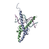
 PDBj
PDBj


 集合体
集合体

 分子量: 18.015 Da / 分子数: 83 / 由来タイプ: 天然 / 式: H2O
 試料調製
試料調製 解析
解析