 Movie
Movie Controller

[English] 日本語
 Yorodumi
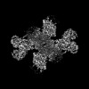
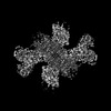
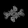
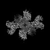
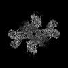
- PDB-7wyw: Cryo-EM structure of Na+,K+-ATPase in the E2P state formed by ino... -
+ Open data

- Basic information
| Entry | Database: PDB / ID: 7wyw | |||||||||||||||||||||
|---|---|---|---|---|---|---|---|---|---|---|---|---|---|---|---|---|---|---|---|---|---|---|
| Title | Cryo-EM structure of Na+,K+-ATPase in the E2P state formed by inorganic phosphate | |||||||||||||||||||||
 Components Components |
| |||||||||||||||||||||
 Keywords Keywords | MEMBRANE PROTEIN / Na+ / K+-ATPase / ion transport / TRANSPORT PROTEIN | |||||||||||||||||||||
| Function / homology |  Function and homology information Function and homology informationregulation of monoatomic ion transport / P-type sodium:potassium-exchanging transporter activity / sodium:potassium-exchanging ATPase complex / sodium ion export across plasma membrane / intracellular sodium ion homeostasis / ATPase activator activity / potassium ion import across plasma membrane / intracellular potassium ion homeostasis / sodium channel regulator activity / monoatomic ion transport ...regulation of monoatomic ion transport / P-type sodium:potassium-exchanging transporter activity / sodium:potassium-exchanging ATPase complex / sodium ion export across plasma membrane / intracellular sodium ion homeostasis / ATPase activator activity / potassium ion import across plasma membrane / intracellular potassium ion homeostasis / sodium channel regulator activity / monoatomic ion transport / proton transmembrane transport / ATP hydrolysis activity / ATP binding / membrane / metal ion binding / plasma membrane Similarity search - Function | |||||||||||||||||||||
| Biological species |  Squalus acanthias (spiny dogfish) Squalus acanthias (spiny dogfish) | |||||||||||||||||||||
| Method | ELECTRON MICROSCOPY / single particle reconstruction / cryo EM / Resolution: 3.8 Å | |||||||||||||||||||||
 Authors Authors | Kanai, R. / Cornelius, F. / Vilsen, B. / Toyoshima, C. | |||||||||||||||||||||
| Funding support |  Japan, Japan,  Denmark, 6items Denmark, 6items
| |||||||||||||||||||||
 Citation Citation | Journal: Proc Natl Acad Sci U S A / Year: 2022 Title: Cryoelectron microscopy of Na,K-ATPase in the two E2P states with and without cardiotonic steroids. Authors: Ryuta Kanai / Flemming Cornelius / Bente Vilsen / Chikashi Toyoshima / Abstract: Cryoelectron microscopy (cryo-EM) was applied to Na+,K+-ATPase (NKA) to determine the structures of two E2P states, one (E2PATP) formed by ATP and Mg2+ in the forward reaction, and the other (E2PPi) ...Cryoelectron microscopy (cryo-EM) was applied to Na+,K+-ATPase (NKA) to determine the structures of two E2P states, one (E2PATP) formed by ATP and Mg2+ in the forward reaction, and the other (E2PPi) formed by inorganic phosphate (Pi) and Mg2+ in the backward reaction, with and without ouabain or istaroxime, representatives of classical and new-generation cardiotonic steroids (CTSs). These two E2P states exhibit different biochemical properties. In particular, K+-sensitive acceleration of the dephosphorylation reaction is not observed with E2PPi, attributed to the presence of a Mg2+ ion in the transmembrane cation binding sites. The cryo-EM structures of NKA demonstrate that the two E2P structures are nearly identical but Mg2+ in the transmembrane binding cavity is identified only in E2PPi, corroborating the idea that it should be denoted as E2PPi·Mg2+. We can now explain why the absence of transmembrane Mg2+ in E2PATP confers the K+ sensitivity in dephosphorylation. In addition, we show that ATP bridges the actuator (A) and nucleotide binding (N) domains, stabilizing the E2PATP state; CTS binding causes hardly any changes in the structure of NKA, both in E2PATP and E2PPi·Mg2+, indicating that the binding mechanism is conformational selection; and istaroxime binds to NKA, extending its aminoalkyloxime group deep into the cation binding site. This orientation is upside down compared to that of classical CTSs with respect to the steroid ring. Notably, mobile parts of NKA are resolved substantially better in the electron microscopy (EM) maps than in previous X-ray structures, including sugars sticking out from the β-subunit and many phospholipid molecules. | |||||||||||||||||||||
| History |
|
- Structure visualization
| Structure viewer | Molecule: MolmilJmol/JSmol |
|---|
- Downloads & links
-Download
| PDBx/mmCIF format | 7wyw.cif.gz | 528.8 KB | Display | PDBx/mmCIF format |
|---|---|---|---|---|
| PDB format | pdb7wyw.ent.gz | 417.9 KB | Display | PDB format |
| PDBx/mmJSON format | 7wyw.json.gz | Tree view | PDBx/mmJSON format | |
| Others |  Other downloads Other downloads |
-Validation report
| Arichive directory | https://data.pdbj.org/pub/pdb/validation_reports/wy/7wywftp://data.pdbj.org/pub/pdb/validation_reports/wy/7wyw | HTTPS FTP |
|---|
-Related structure data
| Related structure data | 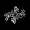 32896MC  7wysC  7wytC 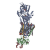 7wyuC 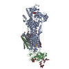 7wyvC  7wyxC  7wyyC  7wyzC  7wz0C C: citing same article ( M: map data used to model this data |
|---|---|
| Similar structure data |

-Links
 PDBj
PDBj


- Assembly
| Deposited unit | 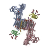
|
|---|---|
| 1 | 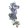
|
| 2 | 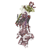
|
| Noncrystallographic symmetry (NCS) | NCS oper: (Code: givenMatrix: (-0.999998107648, -0.00135798468161, -0.00139304644749), (0.00135677555218, -0.999998702387, 0.00086855308711), (-0.00139422412164, 0.000866661392139, 0.999998652518) ...NCS oper: (Code: given Matrix: (-0.999998107648, -0.00135798468161, -0.00139304644749), Vector: |
-Components
-Protein , 3 types, 6 molecules ACBDGE
| #1: Protein | Mass: 113389.859 Da / Num. of mol.: 2 / Source method: isolated from a natural source / Source: (natural) Squalus acanthias (spiny dogfish) / References: UniProt: Q4H132#2: Protein | Mass: 35176.125 Da / Num. of mol.: 2 / Source method: isolated from a natural source / Source: (natural) Squalus acanthias (spiny dogfish) / References: UniProt: C4IX13#3: Protein | Mass: 10195.847 Da / Num. of mol.: 2 / Source method: isolated from a natural source / Source: (natural) Squalus acanthias (spiny dogfish) / References: UniProt: Q70Q12 |
|---|
-Sugars , 4 types, 8 molecules
| #4: Polysaccharide | | #5: Polysaccharide | #6: Polysaccharide | #7: Polysaccharide | |
|---|
-Non-polymers , 4 types, 48 molecules 


| #8: Chemical | ChemComp-MG /  Mass: 24.305 Da / Num. of mol.: 6 / Source method: isolated from a natural source / Formula: Mg Mass: 24.305 Da / Num. of mol.: 6 / Source method: isolated from a natural source / Formula: Mg#9: Chemical | ChemComp-CLR /  Mass: 386.654 Da / Num. of mol.: 8 / Source method: obtained synthetically / Formula: C27H46O Mass: 386.654 Da / Num. of mol.: 8 / Source method: obtained synthetically / Formula: C27H46O#10: Chemical | ChemComp-PCW /  Mass: 787.121 Da / Num. of mol.: 24 / Source method: obtained synthetically / Formula: C44H85NO8P / Comment: DOPC, phospholipid*YM Mass: 787.121 Da / Num. of mol.: 24 / Source method: obtained synthetically / Formula: C44H85NO8P / Comment: DOPC, phospholipid*YM#11: Water | ChemComp-HOH / | Mass: 18.015 Da / Num. of mol.: 10 / Source method: isolated from a natural source / Formula: H2O |
|---|
-Details
| Has ligand of interest | N |
|---|---|
| Has protein modification | Y |
-Experimental details
-Experiment
| Experiment | Method: ELECTRON MICROSCOPY |
|---|---|
| EM experiment | Aggregation state: PARTICLE / 3D reconstruction method: single particle reconstruction |
- Sample preparation
| Component | Name: Sodium/potassium-transporting ATPase / Type: COMPLEX / Entity ID: #1-#3 / Source: NATURAL | |||||||||||||||||||||||||||||||||||
|---|---|---|---|---|---|---|---|---|---|---|---|---|---|---|---|---|---|---|---|---|---|---|---|---|---|---|---|---|---|---|---|---|---|---|---|---|
| Molecular weight | Experimental value: NO | |||||||||||||||||||||||||||||||||||
| Source (natural) | Organism: Squalus acanthias (spiny dogfish) | |||||||||||||||||||||||||||||||||||
| Buffer solution | pH: 7.5 | |||||||||||||||||||||||||||||||||||
| Buffer component |
| |||||||||||||||||||||||||||||||||||
| Specimen | Conc.: 1.5 mg/ml / Embedding applied: NO / Shadowing applied: NO / Staining applied: NO / Vitrification applied: YES | |||||||||||||||||||||||||||||||||||
| Specimen support | Grid material: COPPER / Grid mesh size: 300 divisions/in. / Grid type: C-flat-1.2/1.3 | |||||||||||||||||||||||||||||||||||
| Vitrification | Instrument: FEI VITROBOT MARK IV / Cryogen name: ETHANE / Humidity: 99.9 % / Chamber temperature: 279 K |
- Electron microscopy imaging
| Experimental equipment |  Model: Titan Krios / Image courtesy: FEI Company |
|---|---|
| Microscopy | Model: FEI TITAN KRIOS |
| Electron gun | Electron source:  FIELD EMISSION GUN / Accelerating voltage: 300 kV / Illumination mode: FLOOD BEAM FIELD EMISSION GUN / Accelerating voltage: 300 kV / Illumination mode: FLOOD BEAM |
| Electron lens | Mode: BRIGHT FIELD / Nominal defocus max: 2500 nm / Nominal defocus min: 1000 nm / Cs: 2.7 mm |
| Image recording | Electron dose: 60 e/Å2 / Film or detector model: GATAN K3 (6k x 4k) |
- Processing
| Software |
| ||||||||||||||||||||||||||||||||||||||||
|---|---|---|---|---|---|---|---|---|---|---|---|---|---|---|---|---|---|---|---|---|---|---|---|---|---|---|---|---|---|---|---|---|---|---|---|---|---|---|---|---|---|
| EM software |
| ||||||||||||||||||||||||||||||||||||||||
| CTF correction | Type: PHASE FLIPPING AND AMPLITUDE CORRECTION | ||||||||||||||||||||||||||||||||||||||||
| Symmetry | Point symmetry: C2 (2 fold cyclic) | ||||||||||||||||||||||||||||||||||||||||
| 3D reconstruction | Resolution: 3.8 Å / Resolution method: FSC 0.143 CUT-OFF / Num. of particles: 46883 / Symmetry type: POINT | ||||||||||||||||||||||||||||||||||||||||
| Atomic model building | B value: 49 / Protocol: RIGID BODY FIT / Space: REAL / Target criteria: Correlation Coeeficient | ||||||||||||||||||||||||||||||||||||||||
| Atomic model building | PDB-ID: 7D91 Accession code: 7D91 / Source name: PDB / Type: experimental model | ||||||||||||||||||||||||||||||||||||||||
| Refinement | Cross valid method: NONE Stereochemistry target values: GeoStd + Monomer Library + CDL v1.2 | ||||||||||||||||||||||||||||||||||||||||
| Displacement parameters | Biso mean: 70.12 Å2 | ||||||||||||||||||||||||||||||||||||||||
| Refine LS restraints |
|

