 Movie
Movie Controller


+ Open data

- Basic information
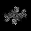
| Entry | Database: PDB / ID: 7wyt | ||||||||||||
|---|---|---|---|---|---|---|---|---|---|---|---|---|---|
| Title | Crystal structures of Na+,K+-ATPase in complex with ouabain | ||||||||||||
 Components Components |
| ||||||||||||
 Keywords Keywords | MEMBRANE PROTEIN / Na+ / K+-ATPase / ion transport / Cardiotonic steroids | ||||||||||||
| Function / homology |  Function and homology information Function and homology information: / Ion homeostasis / Ion transport by P-type ATPases / Na+/K+-exchanging ATPase / regulation of monoatomic ion transport / positive regulation of sodium ion export across plasma membrane / positive regulation of potassium ion import across plasma membrane / P-type sodium:potassium-exchanging transporter activity / sodium ion binding / sodium:potassium-exchanging ATPase complex ...: / Ion homeostasis / Ion transport by P-type ATPases / Na+/K+-exchanging ATPase / regulation of monoatomic ion transport / positive regulation of sodium ion export across plasma membrane / positive regulation of potassium ion import across plasma membrane / P-type sodium:potassium-exchanging transporter activity / sodium ion binding / sodium:potassium-exchanging ATPase complex / membrane repolarization / establishment or maintenance of transmembrane electrochemical gradient / sodium ion export across plasma membrane / regulation of calcium ion transmembrane transport / positive regulation of potassium ion transmembrane transport / ion channel regulator activity / intracellular sodium ion homeostasis / relaxation of cardiac muscle / regulation of cardiac muscle contraction by calcium ion signaling / positive regulation of sodium ion transmembrane transport / organelle membrane / potassium ion binding / potassium ion import across plasma membrane / ATPase activator activity / intracellular potassium ion homeostasis / intercalated disc / lateral plasma membrane / transporter activator activity / sperm flagellum / ATP metabolic process / regulation of sodium ion transport / cardiac muscle contraction / T-tubule / proton transmembrane transport / protein localization to plasma membrane / sarcolemma / transmembrane transport / intracellular calcium ion homeostasis / melanosome / ATPase binding / regulation of gene expression / basolateral plasma membrane / protein-macromolecule adaptor activity / cell adhesion / apical plasma membrane / protein stabilization / innate immune response / axon / protein kinase binding / ATP hydrolysis activity / ATP binding / membrane / plasma membrane Similarity search - Function | ||||||||||||
| Biological species |  | ||||||||||||
| Method |  X-RAY DIFFRACTION / SYNCHROTRON / MOLECULAR REPLACEMENT / Resolution: 2.9 Å X-RAY DIFFRACTION / SYNCHROTRON / MOLECULAR REPLACEMENT / Resolution: 2.9 Å | ||||||||||||
 Authors Authors | Ogawa, H. / Cornelius, F. / Kanai, R. / Motoyama, K. / Vilsen, B. / Toyoshima, C. | ||||||||||||
| Funding support |  Japan, 3items Japan, 3items
| ||||||||||||
 Citation Citation | Journal: Proc Natl Acad Sci U S A / Year: 2022 Title: Cryoelectron microscopy of Na,K-ATPase in the two E2P states with and without cardiotonic steroids. Authors: Ryuta Kanai / Flemming Cornelius / Bente Vilsen / Chikashi Toyoshima /  Abstract: Cryoelectron microscopy (cryo-EM) was applied to Na+,K+-ATPase (NKA) to determine the structures of two E2P states, one (E2PATP) formed by ATP and Mg2+ in the forward reaction, and the other (E2PPi) ...Cryoelectron microscopy (cryo-EM) was applied to Na+,K+-ATPase (NKA) to determine the structures of two E2P states, one (E2PATP) formed by ATP and Mg2+ in the forward reaction, and the other (E2PPi) formed by inorganic phosphate (Pi) and Mg2+ in the backward reaction, with and without ouabain or istaroxime, representatives of classical and new-generation cardiotonic steroids (CTSs). These two E2P states exhibit different biochemical properties. In particular, K+-sensitive acceleration of the dephosphorylation reaction is not observed with E2PPi, attributed to the presence of a Mg2+ ion in the transmembrane cation binding sites. The cryo-EM structures of NKA demonstrate that the two E2P structures are nearly identical but Mg2+ in the transmembrane binding cavity is identified only in E2PPi, corroborating the idea that it should be denoted as E2PPi·Mg2+. We can now explain why the absence of transmembrane Mg2+ in E2PATP confers the K+ sensitivity in dephosphorylation. In addition, we show that ATP bridges the actuator (A) and nucleotide binding (N) domains, stabilizing the E2PATP state; CTS binding causes hardly any changes in the structure of NKA, both in E2PATP and E2PPi·Mg2+, indicating that the binding mechanism is conformational selection; and istaroxime binds to NKA, extending its aminoalkyloxime group deep into the cation binding site. This orientation is upside down compared to that of classical CTSs with respect to the steroid ring. Notably, mobile parts of NKA are resolved substantially better in the electron microscopy (EM) maps than in previous X-ray structures, including sugars sticking out from the β-subunit and many phospholipid molecules. #1: Journal: Proc Natl Acad Sci U S A / Year: 2021Title: Binding of cardiotonic steroids to Na + ,K + -ATPase in the E2P state Authors: Kanai, R. / Cornelius, F. / Ogawa, H. / Motoyama, K. / Vilsen, B. / Toyoshima, C. #2: Journal: To Be PublishedTitle: Cryo-electron microscopy of Na+, K+-ATPase in the two E2P states with and without cardiotonic steroids Authors: Kanai, R. / Cornelius, F. / Vilsen, B. / Toyoshima, C. | ||||||||||||
| History |
|
- Structure visualization
| Structure viewer | Molecule: MolmilJmol/JSmol |
|---|
- Downloads & links
-Download
| PDBx/mmCIF format | 7wyt.cif.gz | 660.5 KB | Display | PDBx/mmCIF format |
|---|---|---|---|---|
| PDB format | pdb7wyt.ent.gz | 434.5 KB | Display | PDB format |
| PDBx/mmJSON format | 7wyt.json.gz | Tree view | PDBx/mmJSON format | |
| Others |  Other downloads Other downloads |
-Validation report
| Arichive directory | https://data.pdbj.org/pub/pdb/validation_reports/wy/7wytftp://data.pdbj.org/pub/pdb/validation_reports/wy/7wyt | HTTPS FTP |
|---|
-Related structure data
| Related structure data |  7wysC 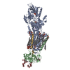 7wyuC 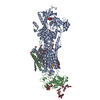 7wyvC 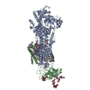 7wywC  7wyxC  7wyyC  7wyzC  7wz0C  6kpu C: citing same article ( S: Starting model for refinement |
|---|---|
| Similar structure data |


-Links
 PDBj
PDBj


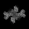
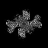
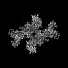
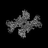
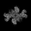
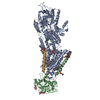
- Assembly
| Deposited unit | 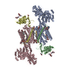
| |||||||||||||||||||||||||||||||||||||||||||||||||||||||||||||||||||||||||||||||||||||||||||||||||||||||||||||||||||||||||||
|---|---|---|---|---|---|---|---|---|---|---|---|---|---|---|---|---|---|---|---|---|---|---|---|---|---|---|---|---|---|---|---|---|---|---|---|---|---|---|---|---|---|---|---|---|---|---|---|---|---|---|---|---|---|---|---|---|---|---|---|---|---|---|---|---|---|---|---|---|---|---|---|---|---|---|---|---|---|---|---|---|---|---|---|---|---|---|---|---|---|---|---|---|---|---|---|---|---|---|---|---|---|---|---|---|---|---|---|---|---|---|---|---|---|---|---|---|---|---|---|---|---|---|---|---|
| 1 | 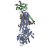
| |||||||||||||||||||||||||||||||||||||||||||||||||||||||||||||||||||||||||||||||||||||||||||||||||||||||||||||||||||||||||||
| 2 | 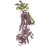
| |||||||||||||||||||||||||||||||||||||||||||||||||||||||||||||||||||||||||||||||||||||||||||||||||||||||||||||||||||||||||||
| Unit cell |
| |||||||||||||||||||||||||||||||||||||||||||||||||||||||||||||||||||||||||||||||||||||||||||||||||||||||||||||||||||||||||||
| Noncrystallographic symmetry (NCS) | NCS domain:
NCS domain segments:
|