 Movie
Movie Controller

[English] 日本語
 Yorodumi
- PDB-7wxe: Crystal Structure of Imine Reductase from Paenibacillus mucilaginosus -
+ Open data

- Basic information
| Entry | Database: PDB / ID: 7wxe | ||||||
|---|---|---|---|---|---|---|---|
| Title | Crystal Structure of Imine Reductase from Paenibacillus mucilaginosus | ||||||
 Components Components | 6-phosphogluconate dehydrogenase | ||||||
 Keywords Keywords | OXIDOREDUCTASE / Imine Reductase | ||||||
| Function / homology |  Function and homology information Function and homology informationtranscription elongation-coupled chromatin remodeling / nucleosome binding / NADP binding / oxidoreductase activity / chromatin / DNA binding Similarity search - Function | ||||||
| Biological species |  Paenibacillus mucilaginosus (bacteria) Paenibacillus mucilaginosus (bacteria) | ||||||
| Method |  X-RAY DIFFRACTION / SYNCHROTRON / MOLECULAR REPLACEMENT / Resolution: 2.5 Å X-RAY DIFFRACTION / SYNCHROTRON / MOLECULAR REPLACEMENT / Resolution: 2.5 Å | ||||||
 Authors Authors | Wu, K. | ||||||
| Funding support |  China, 1items China, 1items
| ||||||
 Citation Citation | Journal: To Be Published Title: Crystal Structure of Imine Reductase from Paenibacillus mucilaginosus Authors: Wu, K. | ||||||
| History |
|
- Structure visualization
| Structure viewer | Molecule: MolmilJmol/JSmol |
|---|
- Downloads & links
-Download
| PDBx/mmCIF format | 7wxe.cif.gz | 132.5 KB | Display | PDBx/mmCIF format |
|---|---|---|---|---|
| PDB format | pdb7wxe.ent.gz | 94.4 KB | Display | PDB format |
| PDBx/mmJSON format | 7wxe.json.gz | Tree view | PDBx/mmJSON format | |
| Others |  Other downloads Other downloads |
-Validation report
| Arichive directory | https://data.pdbj.org/pub/pdb/validation_reports/wx/7wxeftp://data.pdbj.org/pub/pdb/validation_reports/wx/7wxe | HTTPS FTP |
|---|
-Related structure data
| Related structure data | 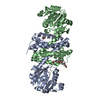 8kfkC 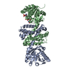 4d3fS S: Starting model for refinement C: citing same article ( |
|---|---|
| Similar structure data |
-Links
 PDBj
PDBj
- Assembly
| Deposited unit | 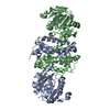
| ||||||||||||
|---|---|---|---|---|---|---|---|---|---|---|---|---|---|
| 1 |
| ||||||||||||
| Unit cell |
| ||||||||||||
| Noncrystallographic symmetry (NCS) | NCS domain:
NCS oper: (Code: givenMatrix: (-0.98327963315, -0.00195843130667, -0.182091536263), (0.0749371649661, -0.91570332468, -0.394806082116), (-0.165968624561, -0.401850203081, 0.900539188456)Vector: 73. ...NCS oper: (Code: given Matrix: (-0.98327963315, -0.00195843130667, -0.182091536263), Vector: |
-Components
| #1: Protein | Mass: 33551.828 Da / Num. of mol.: 2 Source method: isolated from a genetically manipulated source Source: (gene. exp.) Paenibacillus mucilaginosus (bacteria) / Gene: B2K_07400 / Production host: #2: Water | ChemComp-HOH / |  Mass: 18.015 Da / Num. of mol.: 138 / Source method: isolated from a natural source / Formula: H2O Mass: 18.015 Da / Num. of mol.: 138 / Source method: isolated from a natural source / Formula: H2O |
|---|
-Experimental details
-Experiment
| Experiment | Method: X-RAY DIFFRACTION / Number of used crystals: 1 |
|---|
- Sample preparation
| Crystal | Density Matthews: 2.4 Å3/Da / Density % sol: 48.65 % |
|---|---|
| Crystal grow | Temperature: 293 K / Method: vapor diffusion, hanging drop / Details: ammouim sulfate, sodium acetate |
-Data collection
| Diffraction | Mean temperature: 100 K / Serial crystal experiment: N |
|---|---|
| Diffraction source | Source: SYNCHROTRON / Site: SSRF / Beamline: BL18U1 / Wavelength: 0.9792 Å |
| Detector | Type: DECTRIS PILATUS 6M / Detector: PIXEL / Date: Oct 12, 2021 |
| Radiation | Protocol: SINGLE WAVELENGTH / Monochromatic (M) / Laue (L): M / Scattering type: x-ray |
| Radiation wavelength | Wavelength: 0.9792 Å / Relative weight: 1 |
| Reflection | Resolution: 2.5→50 Å / Num. obs: 43954 / % possible obs: 99.8 % / Redundancy: 12.6 % / Biso Wilson estimate: 28.8 Å2 / Rmerge(I) obs: 0.127 / Rpim(I) all: 0.043 / Net I/σ(I): 12.8 |
| Reflection shell | Resolution: 2.5→2.54 Å / Redundancy: 11.6 % / Rmerge(I) obs: 0.786 / Mean I/σ(I) obs: 2 / Num. unique obs: 1963 / Rpim(I) all: 0.421 / % possible all: 99.8 |
- Processing
| Software |
| ||||||||||||||||||||||||||||||||||||||||||||||||||||||||||||||||||||||||||||||||||||||||||||||||||||||||||||||||||||||||||||||||||||||||||||||||||||||||||
|---|---|---|---|---|---|---|---|---|---|---|---|---|---|---|---|---|---|---|---|---|---|---|---|---|---|---|---|---|---|---|---|---|---|---|---|---|---|---|---|---|---|---|---|---|---|---|---|---|---|---|---|---|---|---|---|---|---|---|---|---|---|---|---|---|---|---|---|---|---|---|---|---|---|---|---|---|---|---|---|---|---|---|---|---|---|---|---|---|---|---|---|---|---|---|---|---|---|---|---|---|---|---|---|---|---|---|---|---|---|---|---|---|---|---|---|---|---|---|---|---|---|---|---|---|---|---|---|---|---|---|---|---|---|---|---|---|---|---|---|---|---|---|---|---|---|---|---|---|---|---|---|---|---|---|---|
| Refinement | Method to determine structure: MOLECULAR REPLACEMENT Starting model: 4D3F Resolution: 2.5→28.96 Å / SU ML: 0.2369 / Cross valid method: FREE R-VALUE / σ(F): 1.35 / Phase error: 24.3822 Stereochemistry target values: GeoStd + Monomer Library + CDL v1.2
| ||||||||||||||||||||||||||||||||||||||||||||||||||||||||||||||||||||||||||||||||||||||||||||||||||||||||||||||||||||||||||||||||||||||||||||||||||||||||||
| Solvent computation | Shrinkage radii: 0.9 Å / VDW probe radii: 1.11 Å / Solvent model: FLAT BULK SOLVENT MODEL | ||||||||||||||||||||||||||||||||||||||||||||||||||||||||||||||||||||||||||||||||||||||||||||||||||||||||||||||||||||||||||||||||||||||||||||||||||||||||||
| Displacement parameters | Biso mean: 40.52 Å2 | ||||||||||||||||||||||||||||||||||||||||||||||||||||||||||||||||||||||||||||||||||||||||||||||||||||||||||||||||||||||||||||||||||||||||||||||||||||||||||
| Refinement step | Cycle: LAST / Resolution: 2.5→28.96 Å
| ||||||||||||||||||||||||||||||||||||||||||||||||||||||||||||||||||||||||||||||||||||||||||||||||||||||||||||||||||||||||||||||||||||||||||||||||||||||||||
| Refine LS restraints |
| ||||||||||||||||||||||||||||||||||||||||||||||||||||||||||||||||||||||||||||||||||||||||||||||||||||||||||||||||||||||||||||||||||||||||||||||||||||||||||
| Refine LS restraints NCS | Type: Torsion NCS / Rms dev position: 1.29485798619 Å | ||||||||||||||||||||||||||||||||||||||||||||||||||||||||||||||||||||||||||||||||||||||||||||||||||||||||||||||||||||||||||||||||||||||||||||||||||||||||||
| LS refinement shell |
|
