 Movie
Movie Controller


+ Open data

- Basic information
| Entry | Database: PDB / ID: 7wx6 | |||||||||
|---|---|---|---|---|---|---|---|---|---|---|
| Title | A Legionella acetyltransferase VipF | |||||||||
 Components Components | N-acetyltransferase | |||||||||
 Keywords Keywords | TRANSFERASE / Toxin / acetyltransferase | |||||||||
| Function / homology |  Function and homology information Function and homology informationacyltransferase activity, transferring groups other than amino-acyl groups Similarity search - Function | |||||||||
| Biological species |   Legionella pneumophila (bacteria) Legionella pneumophila (bacteria) | |||||||||
| Method |  X-RAY DIFFRACTION / SYNCHROTRON / MOLECULAR REPLACEMENT / Resolution: 2.273 Å X-RAY DIFFRACTION / SYNCHROTRON / MOLECULAR REPLACEMENT / Resolution: 2.273 Å | |||||||||
 Authors Authors | Chen, T.T. / Lin, Y.L. / Chen, Z. / Han, A.D. | |||||||||
| Funding support |  China, 2items China, 2items
| |||||||||
 Citation Citation | Journal: Acta Crystallogr D Struct Biol / Year: 2022 Title: Structural basis for the acetylation mechanism of the Legionella effector VipF. Authors: Chen, T.T. / Lin, Y. / Zhang, S. / Han, A. | |||||||||
| History |
|
- Structure visualization
| Structure viewer | Molecule: MolmilJmol/JSmol |
|---|
- Downloads & links
-Download
| PDBx/mmCIF format | 7wx6.cif.gz | 133.4 KB | Display | PDBx/mmCIF format |
|---|---|---|---|---|
| PDB format | pdb7wx6.ent.gz | 100.3 KB | Display | PDB format |
| PDBx/mmJSON format | 7wx6.json.gz | Tree view | PDBx/mmJSON format | |
| Others |  Other downloads Other downloads |
-Validation report
| Arichive directory | https://data.pdbj.org/pub/pdb/validation_reports/wx/7wx6ftp://data.pdbj.org/pub/pdb/validation_reports/wx/7wx6 | HTTPS FTP |
|---|
-Related structure data
| Related structure data | 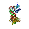 7wx5SC  7wx7C S: Starting model for refinement C: citing same article ( |
|---|---|
| Similar structure data |
-Links
 PDBj
PDBj


- Assembly
| Deposited unit | 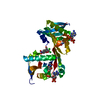
| ||||||||
|---|---|---|---|---|---|---|---|---|---|
| 1 |
| ||||||||
| Unit cell |
| ||||||||
| Components on special symmetry positions |
|
-Components
| #1: Protein | Mass: 35157.297 Da / Num. of mol.: 1 Source method: isolated from a genetically manipulated source Source: (gene. exp.) Legionella pneumophila (bacteria) / Gene: vipF, C3927_15730, DI026_06115 / Production host: | ||||||
|---|---|---|---|---|---|---|---|
| #2: Chemical |   Mass: 767.534 Da / Num. of mol.: 2 / Source method: obtained synthetically / Formula: C21H36N7O16P3S / Feature type: SUBJECT OF INVESTIGATION Mass: 767.534 Da / Num. of mol.: 2 / Source method: obtained synthetically / Formula: C21H36N7O16P3S / Feature type: SUBJECT OF INVESTIGATION#3: Chemical | ChemComp-CLM / | 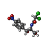  Mass: 323.129 Da / Num. of mol.: 1 / Source method: obtained synthetically / Formula: C11H12Cl2N2O5 / Feature type: SUBJECT OF INVESTIGATION / Comment: antibiotic*YM Mass: 323.129 Da / Num. of mol.: 1 / Source method: obtained synthetically / Formula: C11H12Cl2N2O5 / Feature type: SUBJECT OF INVESTIGATION / Comment: antibiotic*YM#4: Water | ChemComp-HOH / |  Mass: 18.015 Da / Num. of mol.: 105 / Source method: isolated from a natural source / Formula: H2O Mass: 18.015 Da / Num. of mol.: 105 / Source method: isolated from a natural source / Formula: H2OHas ligand of interest | Y | |
-Experimental details
-Experiment
| Experiment | Method: X-RAY DIFFRACTION / Number of used crystals: 1 |
|---|
- Sample preparation
| Crystal | Density Matthews: 2.29 Å3/Da / Density % sol: 46.31 % |
|---|---|
| Crystal grow | Temperature: 298 K / Method: vapor diffusion, sitting drop / Details: PEG8000,EG,HEPES PH7.5 |
-Data collection
| Diffraction | Mean temperature: 293 K / Serial crystal experiment: N |
|---|---|
| Diffraction source | Source: SYNCHROTRON / Site: NFPSS / Beamline: BL19U1 / Wavelength: 0.9793 Å |
| Detector | Type: DECTRIS PILATUS 12M / Detector: PIXEL / Date: Jul 22, 2018 |
| Radiation | Protocol: SINGLE WAVELENGTH / Monochromatic (M) / Laue (L): M / Scattering type: x-ray |
| Radiation wavelength | Wavelength: 0.9793 Å / Relative weight: 1 |
| Reflection | Resolution: 2.273→52.773 Å / Num. obs: 14810 / % possible obs: 97.43 % / Redundancy: 13.1 % / CC1/2: 0.983 / Net I/σ(I): 33.846 |
| Reflection shell | Resolution: 2.273→2.32 Å / Num. unique obs: 14810 / CC1/2: 0.945 |
- Processing
| Software |
| ||||||||||||||||||||||||||||||||||||||||||||||||||||||||||||||||||||||||
|---|---|---|---|---|---|---|---|---|---|---|---|---|---|---|---|---|---|---|---|---|---|---|---|---|---|---|---|---|---|---|---|---|---|---|---|---|---|---|---|---|---|---|---|---|---|---|---|---|---|---|---|---|---|---|---|---|---|---|---|---|---|---|---|---|---|---|---|---|---|---|---|---|---|
| Refinement | Method to determine structure: MOLECULAR REPLACEMENT Starting model: 7WX5 Resolution: 2.273→52.773 Å / SU ML: 0.31 / Cross valid method: THROUGHOUT / σ(F): 0.34 / Phase error: 24.11 / Stereochemistry target values: MLHL
| ||||||||||||||||||||||||||||||||||||||||||||||||||||||||||||||||||||||||
| Solvent computation | Shrinkage radii: 0.9 Å / VDW probe radii: 1.11 Å / Solvent model: FLAT BULK SOLVENT MODEL | ||||||||||||||||||||||||||||||||||||||||||||||||||||||||||||||||||||||||
| Displacement parameters | Biso max: 130.77 Å2 / Biso mean: 33.5335 Å2 / Biso min: 8.7 Å2 | ||||||||||||||||||||||||||||||||||||||||||||||||||||||||||||||||||||||||
| Refinement step | Cycle: final / Resolution: 2.273→52.773 Å
| ||||||||||||||||||||||||||||||||||||||||||||||||||||||||||||||||||||||||
| Refine LS restraints |
| ||||||||||||||||||||||||||||||||||||||||||||||||||||||||||||||||||||||||
| LS refinement shell | Refine-ID: X-RAY DIFFRACTION / Rfactor Rfree error: 0
| ||||||||||||||||||||||||||||||||||||||||||||||||||||||||||||||||||||||||
| Refinement TLS params. | Method: refined / Origin x: -13.3867 Å / Origin y: -15.4362 Å / Origin z: 11.7052 Å
| ||||||||||||||||||||||||||||||||||||||||||||||||||||||||||||||||||||||||
| Refinement TLS group |
|
