 Movie
Movie Controller


+ Open data

- Basic information
| Entry | Database: PDB / ID: 7wu9 | |||||||||||||||||||||
|---|---|---|---|---|---|---|---|---|---|---|---|---|---|---|---|---|---|---|---|---|---|---|
| Title | Cryo-EM structure of the human EP3-Gi signaling complex | |||||||||||||||||||||
 Components Components |
| |||||||||||||||||||||
 Keywords Keywords | SIGNALING PROTEIN / GPCR / signal transduction / MEMBRANE PROTEIN | |||||||||||||||||||||
| Function / homology |  Function and homology information Function and homology informationnegative regulation of gastric acid secretion / intestine smooth muscle contraction / prostaglandin E receptor activity / cell death / Prostanoid ligand receptors / positive regulation of fever generation / adenylate cyclase inhibitor activity / positive regulation of protein localization to cell cortex / T cell migration / positive regulation of relaxation of smooth muscle ...negative regulation of gastric acid secretion / intestine smooth muscle contraction / prostaglandin E receptor activity / cell death / Prostanoid ligand receptors / positive regulation of fever generation / adenylate cyclase inhibitor activity / positive regulation of protein localization to cell cortex / T cell migration / positive regulation of relaxation of smooth muscle / Adenylate cyclase inhibitory pathway / D2 dopamine receptor binding / adenylate cyclase-inhibiting serotonin receptor signaling pathway / G protein-coupled serotonin receptor binding / cellular response to forskolin / regulation of mitotic spindle organization / chemokine-mediated signaling pathway / Regulation of insulin secretion / neuropeptide signaling pathway / response to prostaglandin E / positive regulation of cholesterol biosynthetic process / negative regulation of insulin secretion / G protein-coupled receptor binding / response to peptide hormone / centriolar satellite / G-protein beta/gamma-subunit complex binding / adenylate cyclase-modulating G protein-coupled receptor signaling pathway / adenylate cyclase-inhibiting G protein-coupled receptor signaling pathway / Olfactory Signaling Pathway / Activation of the phototransduction cascade / G protein-coupled acetylcholine receptor signaling pathway / G beta:gamma signalling through PLC beta / Presynaptic function of Kainate receptors / Thromboxane signalling through TP receptor / Activation of G protein gated Potassium channels / Inhibition of voltage gated Ca2+ channels via Gbeta/gamma subunits / G-protein activation / Glucagon signaling in metabolic regulation / Prostacyclin signalling through prostacyclin receptor / G beta:gamma signalling through CDC42 / Synthesis, secretion, and inactivation of Glucagon-like Peptide-1 (GLP-1) / G beta:gamma signalling through BTK / photoreceptor disc membrane / ADP signalling through P2Y purinoceptor 12 / Glucagon-type ligand receptors / GDP binding / Sensory perception of sweet, bitter, and umami (glutamate) taste / Adrenaline,noradrenaline inhibits insulin secretion / nuclear envelope / Vasopressin regulates renal water homeostasis via Aquaporins / Glucagon-like Peptide-1 (GLP1) regulates insulin secretion / G alpha (z) signalling events / cellular response to catecholamine stimulus / ADP signalling through P2Y purinoceptor 1 / ADORA2B mediated anti-inflammatory cytokines production / G beta:gamma signalling through PI3Kgamma / adenylate cyclase-activating dopamine receptor signaling pathway / Cooperation of PDCL (PhLP1) and TRiC/CCT in G-protein beta folding / GPER1 signaling / cellular response to prostaglandin E stimulus / heterotrimeric G-protein complex / G alpha (12/13) signalling events / Inactivation, recovery and regulation of the phototransduction cascade / G-protein beta-subunit binding / extracellular vesicle / sensory perception of taste / sperm principal piece / Thrombin signalling through proteinase activated receptors (PARs) / adenylate cyclase-activating G protein-coupled receptor signaling pathway / signaling receptor complex adaptor activity / positive regulation of cytosolic calcium ion concentration / retina development in camera-type eye / GTPase binding / fibroblast proliferation / G protein activity / midbody / Ca2+ pathway / cell cortex / High laminar flow shear stress activates signaling by PIEZO1 and PECAM1:CDH5:KDR in endothelial cells / G alpha (i) signalling events / G alpha (s) signalling events / G alpha (q) signalling events / phospholipase C-activating G protein-coupled receptor signaling pathway / Hydrolases; Acting on acid anhydrides; Acting on GTP to facilitate cellular and subcellular movement / Ras protein signal transduction / Extra-nuclear estrogen signaling / cell population proliferation / ciliary basal body / G protein-coupled receptor signaling pathway / inflammatory response / cell division / lysosomal membrane / GTPase activity / centrosome / synapse / GTP binding / protein-containing complex binding / nucleolus / magnesium ion binding / Golgi apparatus Similarity search - Function | |||||||||||||||||||||
| Biological species |  Homo sapiens (human) Homo sapiens (human) | |||||||||||||||||||||
| Method | ELECTRON MICROSCOPY / single particle reconstruction / cryo EM / Resolution: 3.375 Å | |||||||||||||||||||||
 Authors Authors | Suno, R. / Sugita, Y. / Morimoto, K. / Iwasaki, K. / Kato, T. / Kobayashi, T. | |||||||||||||||||||||
| Funding support |  Japan, 6items Japan, 6items
| |||||||||||||||||||||
 Citation Citation | Journal: Cell Rep / Year: 2022 Title: Structural insights into the G protein selectivity revealed by the human EP3-G signaling complex. Authors: Ryoji Suno / Yukihiko Sugita / Kazushi Morimoto / Hiroko Takazaki / Hirokazu Tsujimoto / Mika Hirose / Chiyo Suno-Ikeda / Norimichi Nomura / Tomoya Hino / Asuka Inoue / Kenji Iwasaki / ...Authors: Ryoji Suno / Yukihiko Sugita / Kazushi Morimoto / Hiroko Takazaki / Hirokazu Tsujimoto / Mika Hirose / Chiyo Suno-Ikeda / Norimichi Nomura / Tomoya Hino / Asuka Inoue / Kenji Iwasaki / Takayuki Kato / So Iwata / Takuya Kobayashi / Abstract: Prostaglandin receptors have been implicated in a wide range of functions, including inflammation, immune response, reproduction, and cancer. Our group has previously determined the crystal structure ...Prostaglandin receptors have been implicated in a wide range of functions, including inflammation, immune response, reproduction, and cancer. Our group has previously determined the crystal structure of the active-like EP3 bound to its endogenous agonist, prostaglandin E. Here, we present the single-particle cryoelectron microscopy (cryo-EM) structure of the human EP3-G signaling complex at a resolution of 3.4 Å. The structure reveals the binding mode of G to EP3 and the structural changes induced in EP3 by G binding. In addition, we compare the structure of the EP3-G complex with other subtypes of prostaglandin receptors (EP2 and EP4) bound to G that have been previously reported and examine the differences in amino acid composition at the receptor-G protein interface. Mutational analysis reveals that the selectivity of the G protein depends on specific amino acid residues in the second intracellular loop and TM5. | |||||||||||||||||||||
| History |
|
- Structure visualization
| Structure viewer | Molecule: MolmilJmol/JSmol |
|---|
- Downloads & links
-Download
| PDBx/mmCIF format | 7wu9.cif.gz | 197.2 KB | Display | PDBx/mmCIF format |
|---|---|---|---|---|
| PDB format | pdb7wu9.ent.gz | 144.9 KB | Display | PDB format |
| PDBx/mmJSON format | 7wu9.json.gz | Tree view | PDBx/mmJSON format | |
| Others |  Other downloads Other downloads |
-Validation report
| Arichive directory | https://data.pdbj.org/pub/pdb/validation_reports/wu/7wu9ftp://data.pdbj.org/pub/pdb/validation_reports/wu/7wu9 | HTTPS FTP |
|---|
-Related structure data
| Related structure data |  32824MC M: map data used to model this data C: citing same article ( |
|---|---|
| Similar structure data |
-Links
 PDBj
PDBj


- Assembly
| Deposited unit | 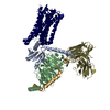
|
|---|---|
| 1 |
|
-Components
| #1: Protein | Mass: 36175.887 Da / Num. of mol.: 1 / Mutation: A173I,V185S,N217Q,S258D,C289L,N308Q Source method: isolated from a genetically manipulated source Source: (gene. exp.) Homo sapiens (human) / Gene: PTGER3 / Production host:   Spodoptera frugiperda (fall armyworm) / References: UniProt: P43115 Spodoptera frugiperda (fall armyworm) / References: UniProt: P43115 |
|---|---|
| #2: Protein | Mass: 40427.969 Da / Num. of mol.: 1 Source method: isolated from a genetically manipulated source Source: (gene. exp.) Homo sapiens (human) / Gene: GNAI1 / Production host: Spodoptera frugiperda (fall armyworm) / References: UniProt: P63096 |
| #3: Protein | Mass: 37728.152 Da / Num. of mol.: 1 Source method: isolated from a genetically manipulated source Source: (gene. exp.) Homo sapiens (human) / Gene: GNB1 / Production host: Spodoptera frugiperda (fall armyworm) / References: UniProt: P62873 |
| #4: Protein | Mass: 7861.143 Da / Num. of mol.: 1 Source method: isolated from a genetically manipulated source Source: (gene. exp.) Homo sapiens (human) / Gene: GNG2 / Production host: Spodoptera frugiperda (fall armyworm) / References: UniProt: P59768 |
| #5: Antibody | Mass: 27318.389 Da / Num. of mol.: 1 Source method: isolated from a genetically manipulated source Source: (gene. exp.)  Brevibacillus agri (bacteria) Brevibacillus agri (bacteria) |
| Has protein modification | Y |
-Experimental details
-Experiment
| Experiment | Method: ELECTRON MICROSCOPY |
|---|---|
| EM experiment | Aggregation state: PARTICLE / 3D reconstruction method: single particle reconstruction |
- Sample preparation
| Component |
| ||||||||||||||||||||||||
|---|---|---|---|---|---|---|---|---|---|---|---|---|---|---|---|---|---|---|---|---|---|---|---|---|---|
| Molecular weight | Experimental value: NO | ||||||||||||||||||||||||
| Source (natural) |
| ||||||||||||||||||||||||
| Source (recombinant) |
| ||||||||||||||||||||||||
| Buffer solution | pH: 7.5 | ||||||||||||||||||||||||
| Specimen | Conc.: 10 mg/ml / Embedding applied: NO / Shadowing applied: NO / Staining applied: NO / Vitrification applied: YES | ||||||||||||||||||||||||
| Vitrification | Cryogen name: ETHANE |
- Electron microscopy imaging
| Experimental equipment |  Model: Titan Krios / Image courtesy: FEI Company |
|---|---|
| Microscopy | Model: FEI TITAN KRIOS |
| Electron gun | Electron source:  FIELD EMISSION GUN / Accelerating voltage: 300 kV / Illumination mode: FLOOD BEAM FIELD EMISSION GUN / Accelerating voltage: 300 kV / Illumination mode: FLOOD BEAM |
| Electron lens | Mode: BRIGHT FIELD / Nominal magnification: 105000 X / Nominal defocus max: 1500 nm / Nominal defocus min: 700 nm / Cs: 0.07 mm / C2 aperture diameter: 50 µm |
| Specimen holder | Cryogen: NITROGEN / Specimen holder model: FEI TITAN KRIOS AUTOGRID HOLDER |
| Image recording | Electron dose: 75 e/Å2 / Film or detector model: GATAN K3 (6k x 4k) |
- Processing
| EM software | Name: SerialEM / Category: image acquisition |
|---|---|
| CTF correction | Type: PHASE FLIPPING AND AMPLITUDE CORRECTION |
| 3D reconstruction | Resolution: 3.375 Å / Resolution method: FSC 0.143 CUT-OFF / Num. of particles: 125572 / Symmetry type: POINT |
