 Movie
Movie Controller

[English] 日本語
 Yorodumi
- PDB-7wu2: Cryo-EM structure of the adhesion GPCR ADGRD1 in complex with miniGs -
+ Open data

- Basic information
| Entry | Database: PDB / ID: 7wu2 | |||||||||||||||||||||||||||||||||
|---|---|---|---|---|---|---|---|---|---|---|---|---|---|---|---|---|---|---|---|---|---|---|---|---|---|---|---|---|---|---|---|---|---|---|
| Title | Cryo-EM structure of the adhesion GPCR ADGRD1 in complex with miniGs | |||||||||||||||||||||||||||||||||
 Components Components |
| |||||||||||||||||||||||||||||||||
 Keywords Keywords | MEMBRANE PROTEIN / Adhesion GPCR / ADGRD1 / G protein / Complex / Signal transduction | |||||||||||||||||||||||||||||||||
| Function / homology |  Function and homology information Function and homology informationregulation of skeletal muscle contraction / G protein-coupled receptor activity / Olfactory Signaling Pathway / Activation of the phototransduction cascade / G protein-coupled acetylcholine receptor signaling pathway / G beta:gamma signalling through PLC beta / Presynaptic function of Kainate receptors / Thromboxane signalling through TP receptor / Activation of G protein gated Potassium channels / Inhibition of voltage gated Ca2+ channels via Gbeta/gamma subunits ...regulation of skeletal muscle contraction / G protein-coupled receptor activity / Olfactory Signaling Pathway / Activation of the phototransduction cascade / G protein-coupled acetylcholine receptor signaling pathway / G beta:gamma signalling through PLC beta / Presynaptic function of Kainate receptors / Thromboxane signalling through TP receptor / Activation of G protein gated Potassium channels / Inhibition of voltage gated Ca2+ channels via Gbeta/gamma subunits / G-protein activation / Glucagon signaling in metabolic regulation / G beta:gamma signalling through CDC42 / Prostacyclin signalling through prostacyclin receptor / Synthesis, secretion, and inactivation of Glucagon-like Peptide-1 (GLP-1) / G beta:gamma signalling through BTK / photoreceptor disc membrane / ADP signalling through P2Y purinoceptor 12 / Glucagon-type ligand receptors / Sensory perception of sweet, bitter, and umami (glutamate) taste / Adrenaline,noradrenaline inhibits insulin secretion / Vasopressin regulates renal water homeostasis via Aquaporins / Glucagon-like Peptide-1 (GLP1) regulates insulin secretion / G alpha (z) signalling events / cellular response to catecholamine stimulus / ADP signalling through P2Y purinoceptor 1 / G beta:gamma signalling through PI3Kgamma / ADORA2B mediated anti-inflammatory cytokines production / adenylate cyclase-activating dopamine receptor signaling pathway / cellular response to prostaglandin E stimulus / Cooperation of PDCL (PhLP1) and TRiC/CCT in G-protein beta folding / GPER1 signaling / heterotrimeric G-protein complex / Inactivation, recovery and regulation of the phototransduction cascade / G alpha (12/13) signalling events / G-protein beta-subunit binding / extracellular vesicle / Thrombin signalling through proteinase activated receptors (PARs) / signaling receptor complex adaptor activity / adenylate cyclase-activating G protein-coupled receptor signaling pathway / GTPase binding / Ca2+ pathway / High laminar flow shear stress activates signaling by PIEZO1 and PECAM1:CDH5:KDR in endothelial cells / G alpha (i) signalling events / G alpha (s) signalling events / G alpha (q) signalling events / Ras protein signal transduction / cell surface receptor signaling pathway / Extra-nuclear estrogen signaling / nuclear speck / G protein-coupled receptor signaling pathway / lysosomal membrane / GTPase activity / synapse / protein-containing complex binding / signal transduction / extracellular exosome / nucleoplasm / membrane / plasma membrane / cytosol / cytoplasm Similarity search - Function | |||||||||||||||||||||||||||||||||
| Biological species |  Homo sapiens (human) Homo sapiens (human) | |||||||||||||||||||||||||||||||||
| Method | ELECTRON MICROSCOPY / single particle reconstruction / cryo EM / Resolution: 2.8 Å | |||||||||||||||||||||||||||||||||
 Authors Authors | Qu, X. / Qiu, N. / Wang, M. / Zhao, Q. / Wu, B. | |||||||||||||||||||||||||||||||||
| Funding support |  China, 5items China, 5items
| |||||||||||||||||||||||||||||||||
 Citation Citation | Journal: Nature / Year: 2022 Title: Structural basis of tethered agonism of the adhesion GPCRs ADGRD1 and ADGRF1. Authors: Xiangli Qu / Na Qiu / Mu Wang / Bingjie Zhang / Juan Du / Zhiwei Zhong / Wei Xu / Xiaojing Chu / Limin Ma / Cuiying Yi / Shuo Han / Wenqing Shui / Qiang Zhao / Beili Wu / Abstract: Adhesion G protein-coupled receptors (aGPCRs) are essential for a variety of physiological processes such as immune responses, organ development, cellular communication, proliferation and homeostasis. ...Adhesion G protein-coupled receptors (aGPCRs) are essential for a variety of physiological processes such as immune responses, organ development, cellular communication, proliferation and homeostasis. An intrinsic manner of activation that involves a tethered agonist in the N-terminal region of the receptor has been proposed for the aGPCRs, but its molecular mechanism remains elusive. Here we report the G protein-bound structures of ADGRD1 and ADGRF1, which exhibit many unique features with regard to the tethered agonism. The stalk region that proceeds the first transmembrane helix acts as the tethered agonist by forming extensive interactions with the transmembrane domain; these interactions are mostly conserved in ADGRD1 and ADGRF1, suggesting that a common stalk-transmembrane domain interaction pattern is shared by members of the aGPCR family. A similar stalk binding mode is observed in the structure of autoproteolysis-deficient ADGRF1, supporting a cleavage-independent manner of receptor activation. The stalk-induced activation is facilitated by a cascade of inter-helix interaction cores that are conserved in positions but show sequence variability in these two aGPCRs. Furthermore, the intracellular region of ADGRF1 contains a specific lipid-binding site, which proves to be functionally important and may serve as the recognition site for the previously discovered endogenous ADGRF1 ligand synaptamide. These findings highlight the diversity and complexity of the signal transduction mechanisms of the aGPCRs. | |||||||||||||||||||||||||||||||||
| History |
|
- Structure visualization
| Structure viewer | Molecule: MolmilJmol/JSmol |
|---|
- Downloads & links
-Download
| PDBx/mmCIF format | 7wu2.cif.gz | 193.5 KB | Display | PDBx/mmCIF format |
|---|---|---|---|---|
| PDB format | pdb7wu2.ent.gz | 145.7 KB | Display | PDB format |
| PDBx/mmJSON format | 7wu2.json.gz | Tree view | PDBx/mmJSON format | |
| Others |  Other downloads Other downloads |
-Validation report
| Arichive directory | https://data.pdbj.org/pub/pdb/validation_reports/wu/7wu2ftp://data.pdbj.org/pub/pdb/validation_reports/wu/7wu2 | HTTPS FTP |
|---|
-Related structure data
| Related structure data | 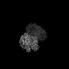 32817MC 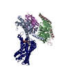 7wu3C 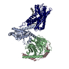 7wu4C  7wu5C M: map data used to model this data C: citing same article ( |
|---|---|
| Similar structure data |
-Links
 PDBj
PDBj


- Assembly
| Deposited unit | 
|
|---|---|
| 1 |
|
-Components
| #1: Protein | Mass: 28387.094 Da / Num. of mol.: 1 Source method: isolated from a genetically manipulated source Source: (gene. exp.) Homo sapiens (human) / Production host:   Spodoptera frugiperda (fall armyworm) Spodoptera frugiperda (fall armyworm) |
|---|---|
| #2: Protein | Mass: 38744.371 Da / Num. of mol.: 1 Source method: isolated from a genetically manipulated source Source: (gene. exp.) Homo sapiens (human) / Gene: GNB1 / Production host: Spodoptera frugiperda (fall armyworm) / References: UniProt: P62873 |
| #3: Protein | Mass: 7861.143 Da / Num. of mol.: 1 Source method: isolated from a genetically manipulated source Source: (gene. exp.) Homo sapiens (human) / Gene: GNG2 / Production host: Spodoptera frugiperda (fall armyworm) / References: UniProt: P59768 |
| #4: Antibody | Mass: 17540.746 Da / Num. of mol.: 1 Source method: isolated from a genetically manipulated source Source: (gene. exp.)  |
| #5: Protein | Mass: 38168.559 Da / Num. of mol.: 1 Source method: isolated from a genetically manipulated source Source: (gene. exp.) Homo sapiens (human) / Gene: ADGRD1, GPR133, PGR25 / Production host: Spodoptera frugiperda (fall armyworm) / References: UniProt: Q6QNK2 |
| Has protein modification | Y |
-Experimental details
-Experiment
| Experiment | Method: ELECTRON MICROSCOPY |
|---|---|
| EM experiment | Aggregation state: PARTICLE / 3D reconstruction method: single particle reconstruction |
- Sample preparation
| Component |
| ||||||||||||||||||||||||
|---|---|---|---|---|---|---|---|---|---|---|---|---|---|---|---|---|---|---|---|---|---|---|---|---|---|
| Source (natural) |
| ||||||||||||||||||||||||
| Source (recombinant) |
| ||||||||||||||||||||||||
| Buffer solution | pH: 7.5 | ||||||||||||||||||||||||
| Specimen | Embedding applied: NO / Shadowing applied: NO / Staining applied: NO / Vitrification applied: YES | ||||||||||||||||||||||||
| Vitrification | Cryogen name: ETHANE |
- Electron microscopy imaging
| Experimental equipment |  Model: Titan Krios / Image courtesy: FEI Company |
|---|---|
| Microscopy | Model: FEI TITAN KRIOS |
| Electron gun | Electron source:  FIELD EMISSION GUN / Accelerating voltage: 300 kV / Illumination mode: FLOOD BEAM FIELD EMISSION GUN / Accelerating voltage: 300 kV / Illumination mode: FLOOD BEAM |
| Electron lens | Mode: BRIGHT FIELD / Nominal defocus max: 1500 nm / Nominal defocus min: 800 nm / Cs: 2.7 mm |
| Image recording | Electron dose: 70 e/Å2 / Film or detector model: GATAN K3 (6k x 4k) |
- Processing
| Software |
| ||||||||||||||||||||||||
|---|---|---|---|---|---|---|---|---|---|---|---|---|---|---|---|---|---|---|---|---|---|---|---|---|---|
| EM software | Name: PHENIX / Category: model refinement | ||||||||||||||||||||||||
| CTF correction | Type: PHASE FLIPPING AND AMPLITUDE CORRECTION | ||||||||||||||||||||||||
| 3D reconstruction | Resolution: 2.8 Å / Resolution method: FSC 0.143 CUT-OFF / Num. of particles: 1266674 / Symmetry type: POINT | ||||||||||||||||||||||||
| Refinement | Cross valid method: NONE Stereochemistry target values: GeoStd + Monomer Library + CDL v1.2 | ||||||||||||||||||||||||
| Displacement parameters | Biso mean: 45.47 Å2 | ||||||||||||||||||||||||
| Refine LS restraints |
|
