 Movie
Movie Controller


+ Open data

- Basic information
| Entry | Database: PDB / ID: 7wpy | ||||||
|---|---|---|---|---|---|---|---|
| Title | AndA_M119A_N121V variant | ||||||
 Components Components | Dioxygenase andA | ||||||
 Keywords Keywords | OXIDOREDUCTASE / non heme iron 2-oxoglutarate dependent dioxygenase / meroterpenoid | ||||||
| Function / homology |  Function and homology information Function and homology informationOxidoreductases; Acting on paired donors, with incorporation or reduction of molecular oxygen; With 2-oxoglutarate as one donor, and incorporation of one atom of oxygen into each donor / terpenoid biosynthetic process / dioxygenase activity / metal ion binding Similarity search - Function | ||||||
| Biological species |  | ||||||
| Method |  X-RAY DIFFRACTION / SYNCHROTRON / MOLECULAR REPLACEMENT / Resolution: 2.25 Å X-RAY DIFFRACTION / SYNCHROTRON / MOLECULAR REPLACEMENT / Resolution: 2.25 Å | ||||||
 Authors Authors | Mori, T. / Chen, H. / Abe, I. | ||||||
| Funding support | 1items
| ||||||
 Citation Citation | Journal: To Be Published Title: AndA_M119A_N121V variant Authors: Mori, T. / Chen, H. / Abe, I. | ||||||
| History |
|
- Structure visualization
| Structure viewer | Molecule: MolmilJmol/JSmol |
|---|
- Downloads & links
-Download
| PDBx/mmCIF format | 7wpy.cif.gz | 272.5 KB | Display | PDBx/mmCIF format |
|---|---|---|---|---|
| PDB format | pdb7wpy.ent.gz | 174.6 KB | Display | PDB format |
| PDBx/mmJSON format | 7wpy.json.gz | Tree view | PDBx/mmJSON format | |
| Others |  Other downloads Other downloads |
-Validation report
| Summary document | 7wpy_validation.pdf.gz | 1.8 MB | Display | wwPDB validaton report |
|---|---|---|---|---|
| Full document | 7wpy_full_validation.pdf.gz | 1.8 MB | Display | |
| Data in XML | 7wpy_validation.xml.gz | 39.7 KB | Display | |
| Data in CIF | 7wpy_validation.cif.gz | 56.6 KB | Display | |
| Arichive directory | https://data.pdbj.org/pub/pdb/validation_reports/wp/7wpyftp://data.pdbj.org/pub/pdb/validation_reports/wp/7wpy | HTTPS FTP |
-Related structure data
| Related structure data | 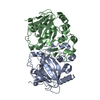 5zm2S S: Starting model for refinement |
|---|---|
| Similar structure data |
-Links
 PDBj
PDBj- Assembly
| Deposited unit | 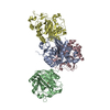
| ||||||||||||
|---|---|---|---|---|---|---|---|---|---|---|---|---|---|
| 1 | 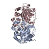
| ||||||||||||
| 2 | 
| ||||||||||||
| Unit cell |
|
-Components
| #1: Protein | Mass: 33641.168 Da / Num. of mol.: 4 Source method: isolated from a genetically manipulated source Source: (gene. exp.)  References: UniProt: A0A097ZPD5, Oxidoreductases; Acting on paired donors, with incorporation or reduction of molecular oxygen; With 2-oxoglutarate as one donor, and incorporation of one atom of oxygen into each donor #2: Chemical | ChemComp-FE /   Mass: 55.845 Da / Num. of mol.: 4 / Source method: obtained synthetically / Formula: Fe / Feature type: SUBJECT OF INVESTIGATION Mass: 55.845 Da / Num. of mol.: 4 / Source method: obtained synthetically / Formula: Fe / Feature type: SUBJECT OF INVESTIGATION#3: Water | ChemComp-HOH / |  Mass: 18.015 Da / Num. of mol.: 375 / Source method: isolated from a natural source / Formula: H2O Mass: 18.015 Da / Num. of mol.: 375 / Source method: isolated from a natural source / Formula: H2OHas ligand of interest | Y | |
|---|
-Experimental details
-Experiment
| Experiment | Method: X-RAY DIFFRACTION / Number of used crystals: 1 |
|---|
- Sample preparation
| Crystal | Density Matthews: 2.13 Å3/Da / Density % sol: 36.52 % |
|---|---|
| Crystal grow | Temperature: 283 K / Method: vapor diffusion, sitting drop / Details: 28% PEG3350, 0.2 M LiCl, 5% DMF |
-Data collection
| Diffraction | Mean temperature: 100 K / Serial crystal experiment: N |
|---|---|
| Diffraction source | Source: SYNCHROTRON / Site: Photon Factory  / Beamline: BL-1A / Wavelength: 1.1 Å / Beamline: BL-1A / Wavelength: 1.1 Å |
| Detector | Type: DECTRIS EIGER X 4M / Detector: PIXEL / Date: Dec 12, 2019 |
| Radiation | Protocol: SINGLE WAVELENGTH / Monochromatic (M) / Laue (L): M / Scattering type: x-ray |
| Radiation wavelength | Wavelength: 1.1 Å / Relative weight: 1 |
| Reflection | Resolution: 2.25→48.74 Å / Num. obs: 48937 / % possible obs: 99.9 % / Redundancy: 3.8 % / Biso Wilson estimate: 33.3 Å2 / CC1/2: 0.996 / Rmerge(I) obs: 0.088 / Net I/σ(I): 8.3 |
| Reflection shell | Resolution: 2.25→2.32 Å / Rmerge(I) obs: 0.539 / Mean I/σ(I) obs: 2.2 / Num. unique obs: 4453 / CC1/2: 0.739 |
- Processing
| Software |
| |||||||||||||||||||||||||||||||||||||||||||||||||||||||||||||||||||||||||||||||||||||||||||||||||||||||||
|---|---|---|---|---|---|---|---|---|---|---|---|---|---|---|---|---|---|---|---|---|---|---|---|---|---|---|---|---|---|---|---|---|---|---|---|---|---|---|---|---|---|---|---|---|---|---|---|---|---|---|---|---|---|---|---|---|---|---|---|---|---|---|---|---|---|---|---|---|---|---|---|---|---|---|---|---|---|---|---|---|---|---|---|---|---|---|---|---|---|---|---|---|---|---|---|---|---|---|---|---|---|---|---|---|---|---|
| Refinement | Method to determine structure: MOLECULAR REPLACEMENT Starting model: 5ZM2 Resolution: 2.25→40.45 Å / SU ML: 0.2293 / Cross valid method: FREE R-VALUE / σ(F): 1.36 / Phase error: 25.8985 Stereochemistry target values: GeoStd + Monomer Library + CDL v1.2
| |||||||||||||||||||||||||||||||||||||||||||||||||||||||||||||||||||||||||||||||||||||||||||||||||||||||||
| Solvent computation | Shrinkage radii: 0.9 Å / VDW probe radii: 1.11 Å / Solvent model: FLAT BULK SOLVENT MODEL | |||||||||||||||||||||||||||||||||||||||||||||||||||||||||||||||||||||||||||||||||||||||||||||||||||||||||
| Displacement parameters | Biso mean: 37.45 Å2 | |||||||||||||||||||||||||||||||||||||||||||||||||||||||||||||||||||||||||||||||||||||||||||||||||||||||||
| Refinement step | Cycle: LAST / Resolution: 2.25→40.45 Å
| |||||||||||||||||||||||||||||||||||||||||||||||||||||||||||||||||||||||||||||||||||||||||||||||||||||||||
| Refine LS restraints |
| |||||||||||||||||||||||||||||||||||||||||||||||||||||||||||||||||||||||||||||||||||||||||||||||||||||||||
| LS refinement shell |
|
