 Movie
Movie Controller


+ Open data

- Basic information
| Entry | Database: PDB / ID: 7wnu | ||||||
|---|---|---|---|---|---|---|---|
| Title | Mycobacterium tuberculosis Rnase J complex with 7nt RNA | ||||||
 Components Components |
| ||||||
 Keywords Keywords | RNA BINDING PROTEIN/RNA / RNA BINDING PROTEIN-RNA COMPLEX | ||||||
| Function / homology |  Function and homology information Function and homology informationRNA exonuclease activity / 5'-3' RNA exonuclease activity / 5'-3' exonuclease activity / RNA endonuclease activity / beta-lactamase activity / beta-lactamase / mRNA processing / rRNA processing / Hydrolases; Acting on ester bonds / RNA binding ...RNA exonuclease activity / 5'-3' RNA exonuclease activity / 5'-3' exonuclease activity / RNA endonuclease activity / beta-lactamase activity / beta-lactamase / mRNA processing / rRNA processing / Hydrolases; Acting on ester bonds / RNA binding / zinc ion binding / cytoplasm Similarity search - Function | ||||||
| Biological species |  Mycobacterium tuberculosis H37Rv (bacteria) Mycobacterium tuberculosis H37Rv (bacteria)synthetic construct (others) | ||||||
| Method |  X-RAY DIFFRACTION / SYNCHROTRON / MOLECULAR REPLACEMENT / Resolution: 3.2 Å X-RAY DIFFRACTION / SYNCHROTRON / MOLECULAR REPLACEMENT / Resolution: 3.2 Å | ||||||
 Authors Authors | Li, J. / Hu, J. / Bao, L. / Zhan, B. / Li, Z. | ||||||
| Funding support |  China, 1items China, 1items
| ||||||
 Citation Citation | Journal: Nat Commun / Year: 2023 Title: Structural insights into RNase J that plays an essential role in Mycobacterium tuberculosis RNA metabolism Authors: Bao, L. / Hu, J. / Zhan, B. / Chi, M. / Li, Z. / Wang, S. / Shan, C. / Zhao, Z. / Guo, Y. / Ding, X. / Ji, C. / Tao, S. / Ni, T. / Zhang, X. / Zhao, G. / Li, J. | ||||||
| History |
|
- Structure visualization
| Structure viewer | Molecule: MolmilJmol/JSmol |
|---|
- Downloads & links
-Download
| PDBx/mmCIF format | 7wnu.cif.gz | 241.1 KB | Display | PDBx/mmCIF format |
|---|---|---|---|---|
| PDB format | pdb7wnu.ent.gz | 176 KB | Display | PDB format |
| PDBx/mmJSON format | 7wnu.json.gz | Tree view | PDBx/mmJSON format | |
| Others |  Other downloads Other downloads |
-Validation report
| Arichive directory | https://data.pdbj.org/pub/pdb/validation_reports/wn/7wnuftp://data.pdbj.org/pub/pdb/validation_reports/wn/7wnu | HTTPS FTP |
|---|
-Related structure data
| Related structure data | 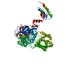 7wntC 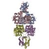 3zq4S S: Starting model for refinement C: citing same article ( |
|---|---|
| Similar structure data |
-Links
 PDBj
PDBj

- Assembly
| Deposited unit | 
| ||||||||||||
|---|---|---|---|---|---|---|---|---|---|---|---|---|---|
| 1 |
| ||||||||||||
| Unit cell |
|
-Components
| #1: Protein | Mass: 59601.332 Da / Num. of mol.: 2 Source method: isolated from a genetically manipulated source Source: (gene. exp.) Mycobacterium tuberculosis H37Rv (bacteria)Strain: H37Rv / Gene: rnj / Production host: #2: RNA chain | Mass: 2259.483 Da / Num. of mol.: 2 / Source method: obtained synthetically / Source: (synth.) synthetic construct (others) #3: Chemical | ChemComp-ZN /   Mass: 65.409 Da / Num. of mol.: 4 / Source method: obtained synthetically / Formula: Zn Mass: 65.409 Da / Num. of mol.: 4 / Source method: obtained synthetically / Formula: ZnHas ligand of interest | N | |
|---|
-Experimental details
-Experiment
| Experiment | Method: X-RAY DIFFRACTION / Number of used crystals: 1 |
|---|
- Sample preparation
| Crystal | Density Matthews: 2.6 Å3/Da / Density % sol: 52.65 % |
|---|---|
| Crystal grow | Temperature: 291.15 K / Method: vapor diffusion, hanging drop Details: 0.2 M Magnesium chloride hexahydrate 0.1 M HEPES pH 7.5 25% w/v Polyethylene glycol 3,350 |
-Data collection
| Diffraction | Mean temperature: 80 K / Serial crystal experiment: N |
|---|---|
| Diffraction source | Source: SYNCHROTRON / Site: SSRF / Beamline: BL19U1 / Wavelength: 0.97853 Å |
| Detector | Type: DECTRIS PILATUS3 6M / Detector: PIXEL / Date: May 28, 2019 |
| Radiation | Protocol: SINGLE WAVELENGTH / Monochromatic (M) / Laue (L): M / Scattering type: x-ray |
| Radiation wavelength | Wavelength: 0.97853 Å / Relative weight: 1 |
| Reflection | Resolution: 3.2→42.88 Å / Num. obs: 21660 / % possible obs: 99.8 % / Redundancy: 2 % / Biso Wilson estimate: 59.25 Å2 / Rmerge(I) obs: 0.0405 / Net I/σ(I): 16.77 |
| Reflection shell | Resolution: 3.2→3.314 Å / Rmerge(I) obs: 0.156 / Num. unique obs: 2157 |
- Processing
| Software |
| |||||||||||||||||||||||||||||||||||||||||||||||||||||||||||||||
|---|---|---|---|---|---|---|---|---|---|---|---|---|---|---|---|---|---|---|---|---|---|---|---|---|---|---|---|---|---|---|---|---|---|---|---|---|---|---|---|---|---|---|---|---|---|---|---|---|---|---|---|---|---|---|---|---|---|---|---|---|---|---|---|---|
| Refinement | Method to determine structure: MOLECULAR REPLACEMENT Starting model: 3ZQ4 Resolution: 3.2→42.88 Å / SU ML: 0.3647 / Cross valid method: FREE R-VALUE / σ(F): 1.34 / Phase error: 27.9492 Stereochemistry target values: GeoStd + Monomer Library + CDL v1.2
| |||||||||||||||||||||||||||||||||||||||||||||||||||||||||||||||
| Solvent computation | Shrinkage radii: 0.9 Å / VDW probe radii: 1.11 Å / Solvent model: FLAT BULK SOLVENT MODEL | |||||||||||||||||||||||||||||||||||||||||||||||||||||||||||||||
| Displacement parameters | Biso mean: 63.03 Å2 | |||||||||||||||||||||||||||||||||||||||||||||||||||||||||||||||
| Refinement step | Cycle: LAST / Resolution: 3.2→42.88 Å
| |||||||||||||||||||||||||||||||||||||||||||||||||||||||||||||||
| Refine LS restraints |
| |||||||||||||||||||||||||||||||||||||||||||||||||||||||||||||||
| LS refinement shell |
|
