 Movie
Movie Controller

[English] 日本語
 Yorodumi
- PDB-7wmb: Crystal structure of 2,3-dihydroxybenzoate decarboxylase mutant W... -
+ Open data

- Basic information
| Entry | Database: PDB / ID: 7wmb | ||||||
|---|---|---|---|---|---|---|---|
| Title | Crystal structure of 2,3-dihydroxybenzoate decarboxylase mutant W23Y from Aspergillus oryzae in complex with catechol | ||||||
 Components Components | Amidohydrolase 2 | ||||||
 Keywords Keywords | HYDROLASE / dihydroxybenzoate decarboxylase | ||||||
| Function / homology |  Function and homology information Function and homology informationsecondary metabolic process / carboxy-lyase activity / hydrolase activity / cytosol Similarity search - Function | ||||||
| Biological species |  | ||||||
| Method |  X-RAY DIFFRACTION / SYNCHROTRON / MOLECULAR REPLACEMENT / Resolution: 2.2 Å X-RAY DIFFRACTION / SYNCHROTRON / MOLECULAR REPLACEMENT / Resolution: 2.2 Å | ||||||
 Authors Authors | Fan, Y. / Xue, S. | ||||||
| Funding support |  China, 1items China, 1items
| ||||||
 Citation Citation | Journal: Appl.Microbiol.Biotechnol. / Year: 2023 Title: The catalytic mechanism of direction-dependent interactions for 2,3-dihydroxybenzoate decarboxylase Authors: Fan, Y. / Wu, S. / Shi, J. / Li, X. / Yang, Y. / Feng, Y. / Xue, S. | ||||||
| History |
|
- Structure visualization
| Structure viewer | Molecule: MolmilJmol/JSmol |
|---|
- Downloads & links
-Download
| PDBx/mmCIF format | 7wmb.cif.gz | 192.5 KB | Display | PDBx/mmCIF format |
|---|---|---|---|---|
| PDB format | pdb7wmb.ent.gz | 120.7 KB | Display | PDB format |
| PDBx/mmJSON format | 7wmb.json.gz | Tree view | PDBx/mmJSON format | |
| Others |  Other downloads Other downloads |
-Validation report
| Arichive directory | https://data.pdbj.org/pub/pdb/validation_reports/wm/7wmbftp://data.pdbj.org/pub/pdb/validation_reports/wm/7wmb | HTTPS FTP |
|---|
-Related structure data
| Related structure data |  7wklC  7wkmC  2dvuS S: Starting model for refinement C: citing same article ( |
|---|---|
| Similar structure data |
-Links
 PDBj
PDBj


- Assembly
| Deposited unit | 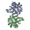
| ||||||||||||
|---|---|---|---|---|---|---|---|---|---|---|---|---|---|
| 1 | 
| ||||||||||||
| Unit cell |
|
-Components
| #1: Protein | Mass: 39020.969 Da / Num. of mol.: 2 / Mutation: W23Y Source method: isolated from a genetically manipulated source Source: (gene. exp.)  #2: Chemical |   Mass: 24.305 Da / Num. of mol.: 2 / Source method: obtained synthetically / Formula: Mg / Feature type: SUBJECT OF INVESTIGATION Mass: 24.305 Da / Num. of mol.: 2 / Source method: obtained synthetically / Formula: Mg / Feature type: SUBJECT OF INVESTIGATION#3: Chemical | 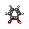  Mass: 110.111 Da / Num. of mol.: 2 / Source method: obtained synthetically / Formula: C6H6O2 / Feature type: SUBJECT OF INVESTIGATION Mass: 110.111 Da / Num. of mol.: 2 / Source method: obtained synthetically / Formula: C6H6O2 / Feature type: SUBJECT OF INVESTIGATION#4: Water | ChemComp-HOH / |  Mass: 18.015 Da / Num. of mol.: 430 / Source method: isolated from a natural source / Formula: H2O Mass: 18.015 Da / Num. of mol.: 430 / Source method: isolated from a natural source / Formula: H2OHas ligand of interest | Y | |
|---|
-Experimental details
-Experiment
| Experiment | Method: X-RAY DIFFRACTION / Number of used crystals: 1 |
|---|
- Sample preparation
| Crystal | Density Matthews: 2.07 Å3/Da / Density % sol: 40.44 % |
|---|---|
| Crystal grow | Temperature: 300 K / Method: vapor diffusion, hanging drop / pH: 5.5 Details: 0.2 M Sodium Chloride, 0.1 M Bis-Tris pH 5.5, 25% w/v PEG 3350 |
-Data collection
| Diffraction | Mean temperature: 100 K / Serial crystal experiment: N |
|---|---|
| Diffraction source | Source: SYNCHROTRON / Site: SSRF / Beamline: BL18U1 / Wavelength: 0.9792 Å |
| Detector | Type: MAR CCD 130 mm / Detector: CCD / Date: Nov 8, 2019 |
| Radiation | Protocol: SINGLE WAVELENGTH / Monochromatic (M) / Laue (L): M / Scattering type: x-ray |
| Radiation wavelength | Wavelength: 0.9792 Å / Relative weight: 1 |
| Reflection | Resolution: 2.19→36 Å / Num. obs: 33777 / % possible obs: 99.52 % / Redundancy: 13.1 % / Biso Wilson estimate: 27.13 Å2 / Rmerge(I) obs: 0.156 / Rrim(I) all: 0.162 / Net I/σ(I): 5.3 |
| Reflection shell | Resolution: 2.19→2.27 Å / Rmerge(I) obs: 0.501 / Mean I/σ(I) obs: 4.22 / Num. unique obs: 6298 / Rrim(I) all: 0.523 |
- Processing
| Software |
| |||||||||||||||||||||||||||||||||||||||||||||||||||||||||||||||||||||||||||||||||||||||||||
|---|---|---|---|---|---|---|---|---|---|---|---|---|---|---|---|---|---|---|---|---|---|---|---|---|---|---|---|---|---|---|---|---|---|---|---|---|---|---|---|---|---|---|---|---|---|---|---|---|---|---|---|---|---|---|---|---|---|---|---|---|---|---|---|---|---|---|---|---|---|---|---|---|---|---|---|---|---|---|---|---|---|---|---|---|---|---|---|---|---|---|---|---|
| Refinement | Method to determine structure: MOLECULAR REPLACEMENT Starting model: 2DVU Resolution: 2.2→35.25 Å / SU ML: 0.222 / Cross valid method: FREE R-VALUE / σ(F): 1.34 / Phase error: 21.0511 Stereochemistry target values: GeoStd + Monomer Library + CDL v1.2
| |||||||||||||||||||||||||||||||||||||||||||||||||||||||||||||||||||||||||||||||||||||||||||
| Solvent computation | Shrinkage radii: 0.9 Å / VDW probe radii: 1.11 Å / Solvent model: FLAT BULK SOLVENT MODEL | |||||||||||||||||||||||||||||||||||||||||||||||||||||||||||||||||||||||||||||||||||||||||||
| Displacement parameters | Biso mean: 25.76 Å2 | |||||||||||||||||||||||||||||||||||||||||||||||||||||||||||||||||||||||||||||||||||||||||||
| Refinement step | Cycle: LAST / Resolution: 2.2→35.25 Å
| |||||||||||||||||||||||||||||||||||||||||||||||||||||||||||||||||||||||||||||||||||||||||||
| Refine LS restraints |
| |||||||||||||||||||||||||||||||||||||||||||||||||||||||||||||||||||||||||||||||||||||||||||
| LS refinement shell |
|
