 Movie
Movie Controller

[English] 日本語
 Yorodumi
- PDB-7wik: Crystal structure of oligoribonuclease of Mycobacterium smegmatis... -
+ Open data

- Basic information
| Entry | Database: PDB / ID: 7wik | ||||||
|---|---|---|---|---|---|---|---|
| Title | Crystal structure of oligoribonuclease of Mycobacterium smegmatis mc2 155 | ||||||
 Components Components | Oligoribonuclease | ||||||
 Keywords Keywords | HYDROLASE / Oligoribonuclease / ORN / RNase H-like fold / DnaQ like exoribonuclease | ||||||
| Function / homology |  Function and homology information Function and homology information3'-5'-RNA exonuclease activity / nucleic acid binding / Hydrolases; Acting on ester bonds / metal ion binding / cytoplasm Similarity search - Function | ||||||
| Biological species |  Mycolicibacterium smegmatis MC2 155 (bacteria) Mycolicibacterium smegmatis MC2 155 (bacteria) | ||||||
| Method |  X-RAY DIFFRACTION / SYNCHROTRON / MOLECULAR REPLACEMENT / Resolution: 1.87 Å X-RAY DIFFRACTION / SYNCHROTRON / MOLECULAR REPLACEMENT / Resolution: 1.87 Å | ||||||
 Authors Authors | Badhwar, P. / Taneja, B. | ||||||
| Funding support | 1items
| ||||||
 Citation Citation | Journal: J.Biol.Chem. / Year: 2022 Title: Three-dimensional structure of a mycobacterial oligoribonuclease reveals a unique C-terminal tail that stabilizes the homodimer. Authors: Badhwar, P. / Khan, S.H. / Taneja, B. | ||||||
| History |
|
- Structure visualization
| Structure viewer | Molecule: MolmilJmol/JSmol |
|---|
- Downloads & links
-Download
| PDBx/mmCIF format | 7wik.cif.gz | 190.6 KB | Display | PDBx/mmCIF format |
|---|---|---|---|---|
| PDB format | pdb7wik.ent.gz | 149.8 KB | Display | PDB format |
| PDBx/mmJSON format | 7wik.json.gz | Tree view | PDBx/mmJSON format | |
| Others |  Other downloads Other downloads |
-Validation report
| Summary document | 7wik_validation.pdf.gz | 1.8 MB | Display | wwPDB validaton report |
|---|---|---|---|---|
| Full document | 7wik_full_validation.pdf.gz | 1.8 MB | Display | |
| Data in XML | 7wik_validation.xml.gz | 38.3 KB | Display | |
| Data in CIF | 7wik_validation.cif.gz | 55.2 KB | Display | |
| Arichive directory | https://data.pdbj.org/pub/pdb/validation_reports/wi/7wikftp://data.pdbj.org/pub/pdb/validation_reports/wi/7wik | HTTPS FTP |
-Related structure data
| Related structure data |  7vh4C 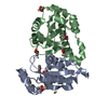 2igiS S: Starting model for refinement C: citing same article ( |
|---|---|
| Similar structure data |
-Links
 PDBj
PDBj- Assembly
| Deposited unit | 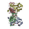
| ||||||||||||||||||||||||||||||||||||||||||||||||||||||||||||||||||||||||||||||||||||||||||||||||||
|---|---|---|---|---|---|---|---|---|---|---|---|---|---|---|---|---|---|---|---|---|---|---|---|---|---|---|---|---|---|---|---|---|---|---|---|---|---|---|---|---|---|---|---|---|---|---|---|---|---|---|---|---|---|---|---|---|---|---|---|---|---|---|---|---|---|---|---|---|---|---|---|---|---|---|---|---|---|---|---|---|---|---|---|---|---|---|---|---|---|---|---|---|---|---|---|---|---|---|---|
| 1 | 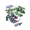
| ||||||||||||||||||||||||||||||||||||||||||||||||||||||||||||||||||||||||||||||||||||||||||||||||||
| 2 | 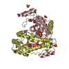
| ||||||||||||||||||||||||||||||||||||||||||||||||||||||||||||||||||||||||||||||||||||||||||||||||||
| Unit cell |
| ||||||||||||||||||||||||||||||||||||||||||||||||||||||||||||||||||||||||||||||||||||||||||||||||||
| Noncrystallographic symmetry (NCS) | NCS domain:
NCS domain segments: Component-ID: _ / Beg auth comp-ID: VAL / Beg label comp-ID: VAL / End auth comp-ID: LEU / End label comp-ID: LEU / Refine code: _ / Auth seq-ID: 1 - 196 / Label seq-ID: 2 - 197
NCS ensembles :
|
-Components
-Protein , 1 types, 4 molecules ABCD
| #1: Protein | Mass: 23257.289 Da / Num. of mol.: 4 Source method: isolated from a genetically manipulated source Source: (gene. exp.) Mycolicibacterium smegmatis MC2 155 (bacteria)Gene: orn / Plasmid: pET28-His10-Smt3 / Production host: |
|---|
-Non-polymers , 7 types, 676 molecules 


| #2: Chemical | ChemComp-K /  Mass: 39.098 Da / Num. of mol.: 4 / Source method: obtained synthetically / Formula: K / Feature type: SUBJECT OF INVESTIGATION Mass: 39.098 Da / Num. of mol.: 4 / Source method: obtained synthetically / Formula: K / Feature type: SUBJECT OF INVESTIGATION#3: Chemical | ChemComp-ACT /  Mass: 59.044 Da / Num. of mol.: 9 / Source method: obtained synthetically / Formula: C2H3O2 Mass: 59.044 Da / Num. of mol.: 9 / Source method: obtained synthetically / Formula: C2H3O2#4: Chemical | ChemComp-SO4 /  Mass: 96.063 Da / Num. of mol.: 13 / Source method: obtained synthetically / Formula: SO4 Mass: 96.063 Da / Num. of mol.: 13 / Source method: obtained synthetically / Formula: SO4#5: Chemical | ChemComp-GOL /  Mass: 92.094 Da / Num. of mol.: 4 / Source method: obtained synthetically / Formula: C3H8O3 Mass: 92.094 Da / Num. of mol.: 4 / Source method: obtained synthetically / Formula: C3H8O3#6: Chemical | ChemComp-MG / |  Mass: 24.305 Da / Num. of mol.: 1 / Source method: isolated from a natural source / Formula: Mg Mass: 24.305 Da / Num. of mol.: 1 / Source method: isolated from a natural source / Formula: Mg#7: Chemical | ChemComp-PEG / |  Mass: 106.120 Da / Num. of mol.: 1 / Source method: obtained synthetically / Formula: C4H10O3 Mass: 106.120 Da / Num. of mol.: 1 / Source method: obtained synthetically / Formula: C4H10O3#8: Water | ChemComp-HOH / | Mass: 18.015 Da / Num. of mol.: 644 / Source method: isolated from a natural source / Formula: H2O |
|---|
-Details
| Has ligand of interest | Y |
|---|
-Experimental details
-Experiment
| Experiment | Method: X-RAY DIFFRACTION / Number of used crystals: 1 |
|---|
- Sample preparation
| Crystal | Density Matthews: 2.32 Å3/Da / Density % sol: 46.97 % / Description: thick, long rods |
|---|---|
| Crystal grow | Temperature: 298 K / Method: vapor diffusion, hanging drop / pH: 8.4 Details: 0.1M Tris-HCl, pH8.4, 0.2M Lithium sulphate, 30% v/v PEG-4000 PH range: 8-9 |
-Data collection
| Diffraction | Mean temperature: 80 K / Serial crystal experiment: N |
|---|---|
| Diffraction source | Source: SYNCHROTRON / Site: ESRF  / Beamline: ID29 / Wavelength: 0.96863 Å / Beamline: ID29 / Wavelength: 0.96863 Å |
| Detector | Type: PSI JUNGFRAU 4M / Detector: PIXEL / Date: May 15, 2018 |
| Radiation | Protocol: SINGLE WAVELENGTH / Monochromatic (M) / Laue (L): M / Scattering type: x-ray |
| Radiation wavelength | Wavelength: 0.96863 Å / Relative weight: 1 |
| Reflection | Resolution: 1.87→81.07 Å / Num. obs: 71290 / % possible obs: 99.5 % / Redundancy: 4.6 % / CC1/2: 0.999 / Rmerge(I) obs: 0.056 / Rpim(I) all: 0.032 / Rrim(I) all: 0.07 / Net I/σ(I): 13.5 |
| Reflection shell | Resolution: 1.874→1.906 Å / Redundancy: 4.3 % / Rmerge(I) obs: 0.694 / Mean I/σ(I) obs: 2 / Num. unique obs: 3483 / CC1/2: 0.803 / Rpim(I) all: 0.377 / Rrim(I) all: 0.794 / % possible all: 99.8 |
- Processing
| Software |
| |||||||||||||||||||||||||||||||||||||||||||||||||||||||||||||||||
|---|---|---|---|---|---|---|---|---|---|---|---|---|---|---|---|---|---|---|---|---|---|---|---|---|---|---|---|---|---|---|---|---|---|---|---|---|---|---|---|---|---|---|---|---|---|---|---|---|---|---|---|---|---|---|---|---|---|---|---|---|---|---|---|---|---|---|
| Refinement | Method to determine structure: MOLECULAR REPLACEMENT Starting model: 2igi Resolution: 1.87→81.07 Å / Cor.coef. Fo:Fc: 0.97 / Cor.coef. Fo:Fc free: 0.952 / SU B: 4.776 / SU ML: 0.128 / Cross valid method: THROUGHOUT / σ(F): 0 / ESU R: 0.146 / ESU R Free: 0.138 / Stereochemistry target values: MAXIMUM LIKELIHOOD Details: HYDROGENS HAVE BEEN ADDED IN THE RIDING POSITIONS U VALUES : REFINED INDIVIDUALLY
| |||||||||||||||||||||||||||||||||||||||||||||||||||||||||||||||||
| Solvent computation | Ion probe radii: 0.8 Å / Shrinkage radii: 0.8 Å / VDW probe radii: 1.2 Å / Solvent model: MASK | |||||||||||||||||||||||||||||||||||||||||||||||||||||||||||||||||
| Displacement parameters | Biso max: 97.89 Å2 / Biso mean: 33.637 Å2 / Biso min: 16.31 Å2
| |||||||||||||||||||||||||||||||||||||||||||||||||||||||||||||||||
| Refinement step | Cycle: final / Resolution: 1.87→81.07 Å
| |||||||||||||||||||||||||||||||||||||||||||||||||||||||||||||||||
| Refine LS restraints |
| |||||||||||||||||||||||||||||||||||||||||||||||||||||||||||||||||
| Refine LS restraints NCS | Refine-ID: X-RAY DIFFRACTION / Type: interatomic distance / Weight position: 0.05
| |||||||||||||||||||||||||||||||||||||||||||||||||||||||||||||||||
| LS refinement shell | Resolution: 1.873→1.922 Å / Rfactor Rfree error: 0 / Total num. of bins used: 20
|
