 Movie
Movie Controller

[English] 日本語
 Yorodumi
- PDB-7wgs: Structure of ClpP from Staphylococcus aureus in complex with (S)-ZG197 -
+ Open data

- Basic information
| Entry | Database: PDB / ID: 7wgs | ||||||||||||
|---|---|---|---|---|---|---|---|---|---|---|---|---|---|
| Title | Structure of ClpP from Staphylococcus aureus in complex with (S)-ZG197 | ||||||||||||
 Components Components | ATP-dependent Clp protease proteolytic subunit | ||||||||||||
 Keywords Keywords | HYDROLASE / ClpP / Staphyloccocus aureus / activator | ||||||||||||
| Function / homology |  Function and homology information Function and homology informationendopeptidase Clp / endopeptidase Clp complex / ATP-dependent peptidase activity / protein quality control for misfolded or incompletely synthesized proteins / ATPase binding / serine-type endopeptidase activity / identical protein binding / cytoplasm Similarity search - Function | ||||||||||||
| Biological species |   Staphylococcus aureus (bacteria) Staphylococcus aureus (bacteria) | ||||||||||||
| Method |  X-RAY DIFFRACTION / SYNCHROTRON / MOLECULAR REPLACEMENT / Resolution: 2.11 Å X-RAY DIFFRACTION / SYNCHROTRON / MOLECULAR REPLACEMENT / Resolution: 2.11 Å | ||||||||||||
 Authors Authors | Yang, C.-G. / Gan, J.H. | ||||||||||||
| Funding support |  China, 3items China, 3items
| ||||||||||||
 Citation Citation | Journal: Nat Commun / Year: 2022 Title: Anti-infective therapy using species-specific activators of Staphylococcus aureus ClpP. Authors: Wei, B. / Zhang, T. / Wang, P. / Pan, Y. / Li, J. / Chen, W. / Zhang, M. / Ji, Q. / Wu, W. / Lan, L. / Gan, J. / Yang, C.G. | ||||||||||||
| History |
|
- Structure visualization
| Structure viewer | Molecule: MolmilJmol/JSmol |
|---|
- Downloads & links
-Download
| PDBx/mmCIF format | 7wgs.cif.gz | 506.3 KB | Display | PDBx/mmCIF format |
|---|---|---|---|---|
| PDB format | pdb7wgs.ent.gz | 419 KB | Display | PDB format |
| PDBx/mmJSON format | 7wgs.json.gz | Tree view | PDBx/mmJSON format | |
| Others |  Other downloads Other downloads |
-Validation report
| Summary document | 7wgs_validation.pdf.gz | 4.5 MB | Display | wwPDB validaton report |
|---|---|---|---|---|
| Full document | 7wgs_full_validation.pdf.gz | 4.6 MB | Display | |
| Data in XML | 7wgs_validation.xml.gz | 96.9 KB | Display | |
| Data in CIF | 7wgs_validation.cif.gz | 126.3 KB | Display | |
| Arichive directory | https://data.pdbj.org/pub/pdb/validation_reports/wg/7wgsftp://data.pdbj.org/pub/pdb/validation_reports/wg/7wgs | HTTPS FTP |
-Related structure data
| Related structure data |  7wh5C  7widC  7xbzC  3staS S: Starting model for refinement C: citing same article ( |
|---|---|
| Similar structure data |
-Links
 PDBj
PDBj- Assembly
| Deposited unit | 
| ||||||||
|---|---|---|---|---|---|---|---|---|---|
| 1 |
| ||||||||
| Unit cell |
|
-Components
| #1: Protein | Mass: 21536.531 Da / Num. of mol.: 14 Source method: isolated from a genetically manipulated source Source: (gene. exp.) Staphylococcus aureus (bacteria) / Gene: ClpP / Production host: #2: Chemical | ChemComp-9A2 / ( 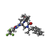  Mass: 532.598 Da / Num. of mol.: 14 / Source method: obtained synthetically / Formula: C28H35F3N4O3 / Feature type: SUBJECT OF INVESTIGATION Mass: 532.598 Da / Num. of mol.: 14 / Source method: obtained synthetically / Formula: C28H35F3N4O3 / Feature type: SUBJECT OF INVESTIGATION#3: Chemical | ChemComp-MPD / (   Mass: 118.174 Da / Num. of mol.: 15 / Source method: obtained synthetically / Formula: C6H14O2 / Comment: precipitant*YM Mass: 118.174 Da / Num. of mol.: 15 / Source method: obtained synthetically / Formula: C6H14O2 / Comment: precipitant*YM#4: Water | ChemComp-HOH / |  Mass: 18.015 Da / Num. of mol.: 739 / Source method: isolated from a natural source / Formula: H2O Mass: 18.015 Da / Num. of mol.: 739 / Source method: isolated from a natural source / Formula: H2OHas ligand of interest | Y | |
|---|
-Experimental details
-Experiment
| Experiment | Method: X-RAY DIFFRACTION / Number of used crystals: 1 |
|---|
- Sample preparation
| Crystal | Density Matthews: 2.84 Å3/Da / Density % sol: 56.76 % |
|---|---|
| Crystal grow | Temperature: 289 K / Method: vapor diffusion, sitting drop Details: 0.2M Sodium chloride, 0.1M Sodium acetate trihydrate pH 4.6, 30% v/v (+/-)-2-Methyl-2,4-pentanediol |
-Data collection
| Diffraction | Mean temperature: 100 K / Serial crystal experiment: N |
|---|---|
| Diffraction source | Source: SYNCHROTRON / Site: SSRF / Beamline: BL02U1 / Wavelength: 0.979 Å |
| Detector | Type: DECTRIS EIGER X 16M / Detector: PIXEL / Date: Oct 6, 2021 |
| Radiation | Protocol: SINGLE WAVELENGTH / Monochromatic (M) / Laue (L): M / Scattering type: x-ray |
| Radiation wavelength | Wavelength: 0.979 Å / Relative weight: 1 |
| Reflection | Resolution: 2.11→145.6 Å / Num. obs: 180268 / % possible obs: 97.6 % / Redundancy: 5.3 % / Rmerge(I) obs: 0.095 / Net I/σ(I): 11.1 |
| Reflection shell | Resolution: 2.11→2.22 Å / Rmerge(I) obs: 0.736 / Num. unique obs: 111 |
- Processing
| Software |
| ||||||||||||||||||||||||||||||||||||||||||||||||||||||||||||
|---|---|---|---|---|---|---|---|---|---|---|---|---|---|---|---|---|---|---|---|---|---|---|---|---|---|---|---|---|---|---|---|---|---|---|---|---|---|---|---|---|---|---|---|---|---|---|---|---|---|---|---|---|---|---|---|---|---|---|---|---|---|
| Refinement | Method to determine structure: MOLECULAR REPLACEMENT Starting model: 3STA Resolution: 2.11→145.6 Å / Cor.coef. Fo:Fc: 0.959 / Cor.coef. Fo:Fc free: 0.953 / SU B: 3.936 / SU ML: 0.103 / Cross valid method: THROUGHOUT / σ(F): 0 / ESU R: 0.188 / ESU R Free: 0.149 / Stereochemistry target values: MAXIMUM LIKELIHOOD Details: HYDROGENS HAVE BEEN ADDED IN THE RIDING POSITIONS U VALUES : REFINED INDIVIDUALLY
| ||||||||||||||||||||||||||||||||||||||||||||||||||||||||||||
| Solvent computation | Ion probe radii: 0.8 Å / Shrinkage radii: 0.8 Å / VDW probe radii: 1.2 Å / Solvent model: MASK | ||||||||||||||||||||||||||||||||||||||||||||||||||||||||||||
| Displacement parameters | Biso max: 122.84 Å2 / Biso mean: 40.685 Å2 / Biso min: 22.12 Å2
| ||||||||||||||||||||||||||||||||||||||||||||||||||||||||||||
| Refinement step | Cycle: final / Resolution: 2.11→145.6 Å
| ||||||||||||||||||||||||||||||||||||||||||||||||||||||||||||
| Refine LS restraints |
| ||||||||||||||||||||||||||||||||||||||||||||||||||||||||||||
| LS refinement shell | Resolution: 2.11→2.16 Å / Rfactor Rfree error: 0
|
