 Movie
Movie Controller

[English] 日本語
 Yorodumi
- PDB-7wgr: Cryo-electron microscopic structure of the 2-oxoglutarate dehydro... -
+ Open data

- Basic information
| Entry | Database: PDB / ID: 7wgr | |||||||||||||||||||||
|---|---|---|---|---|---|---|---|---|---|---|---|---|---|---|---|---|---|---|---|---|---|---|
| Title | Cryo-electron microscopic structure of the 2-oxoglutarate dehydrogenase (E1) component of the human alpha-ketoglutarate (2-oxoglutarate) dehydrogenase complex | |||||||||||||||||||||
 Components Components | 2-oxoglutarate dehydrogenase, mitochondrial | |||||||||||||||||||||
 Keywords Keywords | OXIDOREDUCTASE / Mitochondria / ketoglutarate dehydrogenase / single-particle cryoEM | |||||||||||||||||||||
| Function / homology |  Function and homology information Function and homology informationolfactory bulb mitral cell layer development / OGDH complex synthesizes succinyl-CoA from 2-OG / 2-oxoglutarate decarboxylation to succinyl-CoA / Glycine degradation / oxoglutarate dehydrogenase (succinyl-transferring) / oxoglutarate dehydrogenase (succinyl-transferring) activity / succinyl-CoA metabolic process / tangential migration from the subventricular zone to the olfactory bulb / cerebellar cortex development / oxoglutarate dehydrogenase complex ...olfactory bulb mitral cell layer development / OGDH complex synthesizes succinyl-CoA from 2-OG / 2-oxoglutarate decarboxylation to succinyl-CoA / Glycine degradation / oxoglutarate dehydrogenase (succinyl-transferring) / oxoglutarate dehydrogenase (succinyl-transferring) activity / succinyl-CoA metabolic process / tangential migration from the subventricular zone to the olfactory bulb / cerebellar cortex development / oxoglutarate dehydrogenase complex / 2-oxoglutarate metabolic process / striatum development / pyramidal neuron development / thalamus development / thiamine pyrophosphate binding / tricarboxylic acid cycle / heat shock protein binding / Mitochondrial protein degradation / glycolytic process / hippocampus development / generation of precursor metabolites and energy / mitochondrial membrane / protein-folding chaperone binding / mitochondrial matrix / mitochondrion / metal ion binding / nucleus Similarity search - Function | |||||||||||||||||||||
| Biological species |  Homo sapiens (human) Homo sapiens (human) | |||||||||||||||||||||
| Method | ELECTRON MICROSCOPY / single particle reconstruction / cryo EM / Resolution: 2.92 Å | |||||||||||||||||||||
 Authors Authors | Yu, X. / Yang, W. / Zhong, Y.H. / Ma, X.M. / Gao, Y.Z. | |||||||||||||||||||||
| Funding support |  China, 1items China, 1items
| |||||||||||||||||||||
 Citation Citation | Journal: Biochem.Biophys.Res.Commun. / Year: 2022 Title: Structural basis for the activity and regulation of human alpha-ketoglutarate dehydrogenase revealed by Cryo-EM Authors: Zhong, Y. / Gao, Y. / Zhou, D. / Ma, X. / Chen, H. / Xu, Y. / Yang, W. / Yu, X. | |||||||||||||||||||||
| History |
|
- Structure visualization
| Structure viewer | Molecule: MolmilJmol/JSmol |
|---|
- Downloads & links
-Download
| PDBx/mmCIF format | 7wgr.cif.gz | 312.4 KB | Display | PDBx/mmCIF format |
|---|---|---|---|---|
| PDB format | pdb7wgr.ent.gz | 245.9 KB | Display | PDB format |
| PDBx/mmJSON format | 7wgr.json.gz | Tree view | PDBx/mmJSON format | |
| Others |  Other downloads Other downloads |
-Validation report
| Arichive directory | https://data.pdbj.org/pub/pdb/validation_reports/wg/7wgrftp://data.pdbj.org/pub/pdb/validation_reports/wg/7wgr | HTTPS FTP |
|---|
-Related structure data
| Related structure data |  32485MC M: map data used to model this data C: citing same article ( |
|---|---|
| Similar structure data |
-Links
 PDBj
PDBj


- Assembly
| Deposited unit | 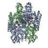
|
|---|---|
| 1 |
|
-Components
| #1: Protein | Mass: 102006.258 Da / Num. of mol.: 2 Source method: isolated from a genetically manipulated source Source: (gene. exp.) Homo sapiens (human) / Gene: OGDH / Production host:  References: UniProt: Q02218, oxoglutarate dehydrogenase (succinyl-transferring) #2: Chemical |   Mass: 40.078 Da / Num. of mol.: 2 / Source method: obtained synthetically / Formula: Ca / Feature type: SUBJECT OF INVESTIGATION Mass: 40.078 Da / Num. of mol.: 2 / Source method: obtained synthetically / Formula: Ca / Feature type: SUBJECT OF INVESTIGATION#3: Chemical |   Mass: 24.305 Da / Num. of mol.: 2 / Source method: obtained synthetically / Formula: Mg / Feature type: SUBJECT OF INVESTIGATION Mass: 24.305 Da / Num. of mol.: 2 / Source method: obtained synthetically / Formula: Mg / Feature type: SUBJECT OF INVESTIGATION#4: Chemical |   Mass: 425.314 Da / Num. of mol.: 2 / Source method: obtained synthetically / Formula: C12H19N4O7P2S / Feature type: SUBJECT OF INVESTIGATION Mass: 425.314 Da / Num. of mol.: 2 / Source method: obtained synthetically / Formula: C12H19N4O7P2S / Feature type: SUBJECT OF INVESTIGATIONHas ligand of interest | Y | Has protein modification | N | |
|---|
-Experimental details
-Experiment
| Experiment | Method: ELECTRON MICROSCOPY |
|---|---|
| EM experiment | Aggregation state: PARTICLE / 3D reconstruction method: single particle reconstruction |
- Sample preparation
| Component | Name: 2-oxoglutarate dehydrogenase, OGDH / Type: COMPLEX / Entity ID: #1 / Source: RECOMBINANT |
|---|---|
| Source (natural) | Organism: Homo sapiens (human) |
| Source (recombinant) | Organism: |
| Buffer solution | pH: 7.4 |
| Specimen | Conc.: 1.5 mg/ml / Embedding applied: NO / Shadowing applied: NO / Staining applied: NO / Vitrification applied: YES |
| Vitrification | Cryogen name: ETHANE-PROPANE |
- Electron microscopy imaging
| Experimental equipment |  Model: Titan Krios / Image courtesy: FEI Company |
|---|---|
| Microscopy | Model: FEI TITAN KRIOS |
| Electron gun | Electron source:  FIELD EMISSION GUN / Accelerating voltage: 300 kV / Illumination mode: FLOOD BEAM FIELD EMISSION GUN / Accelerating voltage: 300 kV / Illumination mode: FLOOD BEAM |
| Electron lens | Mode: BRIGHT FIELD / Nominal defocus max: 2000 nm / Nominal defocus min: 1000 nm |
| Image recording | Electron dose: 60 e/Å2 / Film or detector model: GATAN K2 QUANTUM (4k x 4k) |
- Processing
| Software | Name: PHENIX / Version: 1.19_4092: / Classification: refinement | ||||||||||||||||||||||||
|---|---|---|---|---|---|---|---|---|---|---|---|---|---|---|---|---|---|---|---|---|---|---|---|---|---|
| EM software | Name: PHENIX / Category: model refinement | ||||||||||||||||||||||||
| CTF correction | Type: PHASE FLIPPING AND AMPLITUDE CORRECTION | ||||||||||||||||||||||||
| 3D reconstruction | Resolution: 2.92 Å / Resolution method: FSC 0.143 CUT-OFF / Num. of particles: 266798 / Symmetry type: POINT | ||||||||||||||||||||||||
| Refine LS restraints |
|
