 Movie
Movie Controller

[English] 日本語
 Yorodumi
- PDB-7wbc: Hydroxysteroid dehydrogenase wild-type complexed with NAD+ and (4... -
+ Open data

- Basic information
| Entry | Database: PDB / ID: 7wbc | ||||||
|---|---|---|---|---|---|---|---|
| Title | Hydroxysteroid dehydrogenase wild-type complexed with NAD+ and (4S)-2-2-methyl-2,4-pentanediol | ||||||
 Components Components | SDR family NAD(P)-dependent oxidoreductase | ||||||
 Keywords Keywords | OXIDOREDUCTASE / Hydroxysteroid dehydrogenase / Complex / NAD+ | ||||||
| Function / homology | NICOTINAMIDE-ADENINE-DINUCLEOTIDE / PHOSPHATE ION / :  Function and homology information Function and homology information | ||||||
| Biological species |  Rhodococcus ruber (bacteria) Rhodococcus ruber (bacteria) | ||||||
| Method |  X-RAY DIFFRACTION / SYNCHROTRON / MOLECULAR REPLACEMENT / Resolution: 2 Å X-RAY DIFFRACTION / SYNCHROTRON / MOLECULAR REPLACEMENT / Resolution: 2 Å | ||||||
 Authors Authors | Shi, S.-C. / Zheng, Y.-C. / Xu, J.-H. / Li, C.-X. | ||||||
| Funding support |  China, 1items China, 1items
| ||||||
 Citation Citation | Journal: To Be Published Title: Hydroxysteroid dehydrogenase wild-type complexed with NAD+ and (4S)-2-2-methyl-2,4-pentanediol Authors: Shi, S.-C. / Zheng, Y.-C. / Xu, J.-H. / Li, C.-X. | ||||||
| History |
|
- Structure visualization
| Structure viewer | Molecule: MolmilJmol/JSmol |
|---|
- Downloads & links
-Download
| PDBx/mmCIF format | 7wbc.cif.gz | 118 KB | Display | PDBx/mmCIF format |
|---|---|---|---|---|
| PDB format | pdb7wbc.ent.gz | 88 KB | Display | PDB format |
| PDBx/mmJSON format | 7wbc.json.gz | Tree view | PDBx/mmJSON format | |
| Others |  Other downloads Other downloads |
-Validation report
| Summary document | 7wbc_validation.pdf.gz | 1.1 MB | Display | wwPDB validaton report |
|---|---|---|---|---|
| Full document | 7wbc_full_validation.pdf.gz | 1.1 MB | Display | |
| Data in XML | 7wbc_validation.xml.gz | 23.6 KB | Display | |
| Data in CIF | 7wbc_validation.cif.gz | 33.7 KB | Display | |
| Arichive directory | https://data.pdbj.org/pub/pdb/validation_reports/wb/7wbcftp://data.pdbj.org/pub/pdb/validation_reports/wb/7wbc | HTTPS FTP |
-Related structure data
| Related structure data | 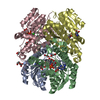 5jy1S S: Starting model for refinement |
|---|---|
| Similar structure data |
-Links
 PDBj
PDBj- Assembly
| Deposited unit | 
| ||||||||||||
|---|---|---|---|---|---|---|---|---|---|---|---|---|---|
| 1 |
| ||||||||||||
| Unit cell |
| ||||||||||||
| Components on special symmetry positions |
|
-Components
-Protein , 1 types, 2 molecules AB
| #1: Protein | Mass: 27430.994 Da / Num. of mol.: 2 Source method: isolated from a genetically manipulated source Source: (gene. exp.) Rhodococcus ruber (bacteria) / Gene: DCN13_20495, F1734_22905 / Production host: |
|---|
-Non-polymers , 6 types, 346 molecules 


| #2: Chemical |  Mass: 118.174 Da / Num. of mol.: 2 / Source method: obtained synthetically / Formula: C6H14O2 / Comment: precipitant*YM Mass: 118.174 Da / Num. of mol.: 2 / Source method: obtained synthetically / Formula: C6H14O2 / Comment: precipitant*YM#3: Chemical |  Mass: 663.425 Da / Num. of mol.: 2 / Source method: obtained synthetically / Formula: C21H27N7O14P2 / Feature type: SUBJECT OF INVESTIGATION / Comment: NAD*YM Mass: 663.425 Da / Num. of mol.: 2 / Source method: obtained synthetically / Formula: C21H27N7O14P2 / Feature type: SUBJECT OF INVESTIGATION / Comment: NAD*YM#4: Chemical | ChemComp-EDO /  Mass: 62.068 Da / Num. of mol.: 4 / Source method: obtained synthetically / Formula: C2H6O2 Mass: 62.068 Da / Num. of mol.: 4 / Source method: obtained synthetically / Formula: C2H6O2#5: Chemical |  Mass: 40.078 Da / Num. of mol.: 2 / Source method: obtained synthetically / Formula: Ca Mass: 40.078 Da / Num. of mol.: 2 / Source method: obtained synthetically / Formula: Ca#6: Chemical | ChemComp-PO4 / |  Mass: 94.971 Da / Num. of mol.: 1 / Source method: obtained synthetically / Formula: PO4 Mass: 94.971 Da / Num. of mol.: 1 / Source method: obtained synthetically / Formula: PO4#7: Water | ChemComp-HOH / | Mass: 18.015 Da / Num. of mol.: 335 / Source method: isolated from a natural source / Formula: H2O |
|---|
-Details
| Has ligand of interest | Y |
|---|
-Experimental details
-Experiment
| Experiment | Method: X-RAY DIFFRACTION / Number of used crystals: 1 |
|---|
- Sample preparation
| Crystal | Density Matthews: 2.11 Å3/Da / Density % sol: 41.71 % |
|---|---|
| Crystal grow | Temperature: 291 K / Method: vapor diffusion, sitting drop Details: 2-(N-Morpholino) ethane sulfonic acid, MgCl2, CaCl2, MPD, P3350, 2-(N-morpholino) ethane sulfonic acid, imidazole |
-Data collection
| Diffraction | Mean temperature: 100 K / Serial crystal experiment: N |
|---|---|
| Diffraction source | Source: SYNCHROTRON / Site: SSRF / Beamline: BL17U1 / Wavelength: 0.9789 Å |
| Detector | Type: DECTRIS PILATUS3 6M / Detector: PIXEL / Date: Jun 9, 2019 |
| Radiation | Protocol: SINGLE WAVELENGTH / Monochromatic (M) / Laue (L): M / Scattering type: x-ray |
| Radiation wavelength | Wavelength: 0.9789 Å / Relative weight: 1 |
| Reflection | Resolution: 2→33.3 Å / Num. obs: 30411 / % possible obs: 95.8 % / Redundancy: 11.2 % / Biso Wilson estimate: 24.42 Å2 / CC1/2: 0.979 / Net I/σ(I): 14.3 |
| Reflection shell | Resolution: 2→2.03 Å / Num. unique obs: 1560 / CC1/2: 0.978 |
- Processing
| Software |
| ||||||||||||||||||||||||||||||||||||||||||||||||||||||||||||||||||||||||
|---|---|---|---|---|---|---|---|---|---|---|---|---|---|---|---|---|---|---|---|---|---|---|---|---|---|---|---|---|---|---|---|---|---|---|---|---|---|---|---|---|---|---|---|---|---|---|---|---|---|---|---|---|---|---|---|---|---|---|---|---|---|---|---|---|---|---|---|---|---|---|---|---|---|
| Refinement | Method to determine structure: MOLECULAR REPLACEMENT Starting model: 5JY1 Resolution: 2→33.3 Å / SU ML: 0.19 / Cross valid method: THROUGHOUT / σ(F): 1.38 / Phase error: 25.4 / Stereochemistry target values: ML
| ||||||||||||||||||||||||||||||||||||||||||||||||||||||||||||||||||||||||
| Solvent computation | Shrinkage radii: 0.9 Å / VDW probe radii: 1.11 Å / Solvent model: FLAT BULK SOLVENT MODEL | ||||||||||||||||||||||||||||||||||||||||||||||||||||||||||||||||||||||||
| Displacement parameters | Biso max: 81.82 Å2 / Biso mean: 26.0029 Å2 / Biso min: 14.25 Å2 | ||||||||||||||||||||||||||||||||||||||||||||||||||||||||||||||||||||||||
| Refinement step | Cycle: final / Resolution: 2→33.3 Å
| ||||||||||||||||||||||||||||||||||||||||||||||||||||||||||||||||||||||||
| LS refinement shell | Refine-ID: X-RAY DIFFRACTION / Rfactor Rfree error: 0
|
