 Movie
Movie Controller

[English] 日本語
 Yorodumi
- PDB-7w8m: Crystal structure of Co-type nitrile hydratase mutant from Pseudo... -
+ Open data

- Basic information
| Entry | Database: PDB / ID: 7w8m | ||||||
|---|---|---|---|---|---|---|---|
| Title | Crystal structure of Co-type nitrile hydratase mutant from Pseudomonas thermophila - A129R | ||||||
 Components Components |
| ||||||
 Keywords Keywords | LYASE / hydratase / catalyze the hydration of nitriles to form amides | ||||||
| Function / homology |  Function and homology information Function and homology informationnitrile catabolic process / nitrile hydratase / nitrile hydratase activity / transition metal ion binding Similarity search - Function | ||||||
| Biological species |  Pseudonocardia thermophila DSM 43832 (bacteria)Pseudonocardia thermophila (bacteria) Pseudonocardia thermophila DSM 43832 (bacteria)Pseudonocardia thermophila (bacteria) | ||||||
| Method |  X-RAY DIFFRACTION / MOLECULAR REPLACEMENT / Resolution: 2.6 Å X-RAY DIFFRACTION / MOLECULAR REPLACEMENT / Resolution: 2.6 Å | ||||||
 Authors Authors | Ma, D. / Cheng, Z.Y. / Hou, X.D. / Peplowski, L. / Lai, Q.P. / Fu, K. / Yin, D.J. / Rao, Y.J. / Zhou, Z.M. | ||||||
| Funding support |  China, 1items China, 1items
| ||||||
 Citation Citation | Journal: Chem Sci / Year: 2022 Title: Insight into the broadened substrate scope of nitrile hydratase by static and dynamic structure analysis. Authors: Ma, D. / Cheng, Z. / Peplowski, L. / Han, L. / Xia, Y. / Hou, X. / Guo, J. / Yin, D. / Rao, Y. / Zhou, Z. #1: Journal: Biochem Biophys Res Commun / Year: 2001Title: Crystal structure of cobalt-containing nitrile hydratase. Authors: Miyanaga, A. / Fushinobu, S. / Ito, K. / Wakagi, T. | ||||||
| History |
|
- Structure visualization
| Structure viewer | Molecule: MolmilJmol/JSmol |
|---|
- Downloads & links
-Download
| PDBx/mmCIF format | 7w8m.cif.gz | 102.5 KB | Display | PDBx/mmCIF format |
|---|---|---|---|---|
| PDB format | pdb7w8m.ent.gz | 76.4 KB | Display | PDB format |
| PDBx/mmJSON format | 7w8m.json.gz | Tree view | PDBx/mmJSON format | |
| Others |  Other downloads Other downloads |
-Validation report
| Arichive directory | https://data.pdbj.org/pub/pdb/validation_reports/w8/7w8mftp://data.pdbj.org/pub/pdb/validation_reports/w8/7w8m | HTTPS FTP |
|---|
-Related structure data
| Related structure data | 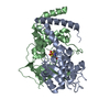 7w8lC 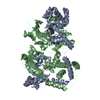 1ireS S: Starting model for refinement C: citing same article ( |
|---|---|
| Similar structure data |
-Links
 PDBj
PDBj


- Assembly
| Deposited unit | 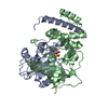
| ||||||||
|---|---|---|---|---|---|---|---|---|---|
| 1 | 
| ||||||||
| Unit cell |
|
-Components
| #1: Protein | Mass: 23290.670 Da / Num. of mol.: 1 Source method: isolated from a genetically manipulated source Source: (gene. exp.) Pseudonocardia thermophila DSM 43832 (bacteria)Gene: SAMN05443637_10361 / Production host: |
|---|---|
| #2: Protein | Mass: 26669.760 Da / Num. of mol.: 1 / Mutation: A129R Source method: isolated from a genetically manipulated source Source: (gene. exp.) Pseudonocardia thermophila (bacteria) / Gene: SAMN05443637_10360 / Production host: |
| #3: Chemical | ChemComp-CO /   Mass: 58.933 Da / Num. of mol.: 1 / Source method: obtained synthetically / Formula: Co / Feature type: SUBJECT OF INVESTIGATION Mass: 58.933 Da / Num. of mol.: 1 / Source method: obtained synthetically / Formula: Co / Feature type: SUBJECT OF INVESTIGATION |
| #4: Water | ChemComp-HOH /  Mass: 18.015 Da / Num. of mol.: 103 / Source method: isolated from a natural source / Formula: H2O Mass: 18.015 Da / Num. of mol.: 103 / Source method: isolated from a natural source / Formula: H2O |
| Has ligand of interest | Y |
| Has protein modification | Y |
-Experimental details
-Experiment
| Experiment | Method: X-RAY DIFFRACTION / Number of used crystals: 1 |
|---|
- Sample preparation
| Crystal | Density Matthews: 2.28 Å3/Da / Density % sol: 46.02 % |
|---|---|
| Crystal grow | Temperature: 289.15 K / Method: vapor diffusion, sitting drop / Details: sodium citrate, HEPES-NaOH, pH 7.5 |
-Data collection
| Diffraction | Mean temperature: 150 K / Serial crystal experiment: N | ||||||||||||||||||||||||||||||||||||||||
|---|---|---|---|---|---|---|---|---|---|---|---|---|---|---|---|---|---|---|---|---|---|---|---|---|---|---|---|---|---|---|---|---|---|---|---|---|---|---|---|---|---|
| Diffraction source | Source: SEALED TUBE / Type: BRUKER D8 QUEST / Wavelength: 1.542 Å | ||||||||||||||||||||||||||||||||||||||||
| Detector | Type: Bruker PHOTON II / Detector: PIXEL / Date: Apr 21, 2021 | ||||||||||||||||||||||||||||||||||||||||
| Radiation | Protocol: SINGLE WAVELENGTH / Monochromatic (M) / Laue (L): M / Scattering type: x-ray | ||||||||||||||||||||||||||||||||||||||||
| Radiation wavelength | Wavelength: 1.542 Å / Relative weight: 1 | ||||||||||||||||||||||||||||||||||||||||
| Reflection | Resolution: 2.598→22.37 Å / Num. obs: 14569 / % possible obs: 96.94 % / Redundancy: 1.82 % / Rsym value: 0.077 / Net I/av σ(I): 12.087 / Net I/σ(I): 12.27 | ||||||||||||||||||||||||||||||||||||||||
| Reflection shell |
|
- Processing
| Software |
| ||||||||||||||||||||||||||||||||||||||||||
|---|---|---|---|---|---|---|---|---|---|---|---|---|---|---|---|---|---|---|---|---|---|---|---|---|---|---|---|---|---|---|---|---|---|---|---|---|---|---|---|---|---|---|---|
| Refinement | Method to determine structure: MOLECULAR REPLACEMENT Starting model: 1IRE Resolution: 2.6→22.37 Å / SU ML: 0.27 / Cross valid method: THROUGHOUT / σ(F): 1.34 / Phase error: 22.1 / Stereochemistry target values: ML
| ||||||||||||||||||||||||||||||||||||||||||
| Solvent computation | Shrinkage radii: 0.9 Å / VDW probe radii: 1.11 Å / Solvent model: FLAT BULK SOLVENT MODEL | ||||||||||||||||||||||||||||||||||||||||||
| Displacement parameters | Biso max: 74.8 Å2 / Biso mean: 17.4282 Å2 / Biso min: 4.42 Å2 | ||||||||||||||||||||||||||||||||||||||||||
| Refinement step | Cycle: final / Resolution: 2.6→22.37 Å
| ||||||||||||||||||||||||||||||||||||||||||
| LS refinement shell | Refine-ID: X-RAY DIFFRACTION / Rfactor Rfree error: 0 / Total num. of bins used: 5
|
