 Movie
Movie Controller


+ Open data

- Basic information
| Entry | Database: PDB / ID: 7w8d | ||||||
|---|---|---|---|---|---|---|---|
| Title | The structure of Deinococcus radiodurans RuvC | ||||||
 Components Components | Crossover junction endodeoxyribonuclease RuvC | ||||||
 Keywords Keywords | HYDROLASE / holliday junction resolvase / DNase / DNA binding | ||||||
| Function / homology |  Function and homology information Function and homology informationcrossover junction endodeoxyribonuclease / Holliday junction resolvase complex / crossover junction DNA endonuclease activity / DNA recombination / DNA repair / magnesium ion binding / DNA binding / cytoplasm Similarity search - Function | ||||||
| Biological species |  Deinococcus radiodurans (radioresistant) Deinococcus radiodurans (radioresistant) | ||||||
| Method |  X-RAY DIFFRACTION / SYNCHROTRON / MOLECULAR REPLACEMENT / Resolution: 2.75016129042 Å X-RAY DIFFRACTION / SYNCHROTRON / MOLECULAR REPLACEMENT / Resolution: 2.75016129042 Å | ||||||
 Authors Authors | Cheng, K. | ||||||
| Funding support |  China, 1items China, 1items
| ||||||
 Citation Citation | Journal: Mbio / Year: 2022 Title: Biochemical and Structural Study of RuvC and YqgF from Deinococcus radiodurans. Authors: Sun, Y. / Yang, J. / Xu, G. / Cheng, K. | ||||||
| History |
|
- Structure visualization
| Structure viewer | Molecule: MolmilJmol/JSmol |
|---|
- Downloads & links
-Download
| PDBx/mmCIF format | 7w8d.cif.gz | 156.1 KB | Display | PDBx/mmCIF format |
|---|---|---|---|---|
| PDB format | pdb7w8d.ent.gz | 102.7 KB | Display | PDB format |
| PDBx/mmJSON format | 7w8d.json.gz | Tree view | PDBx/mmJSON format | |
| Others |  Other downloads Other downloads |
-Validation report
| Arichive directory | https://data.pdbj.org/pub/pdb/validation_reports/w8/7w8dftp://data.pdbj.org/pub/pdb/validation_reports/w8/7w8d | HTTPS FTP |
|---|
-Related structure data
| Related structure data |  7w89C  4ep4S S: Starting model for refinement C: citing same article ( |
|---|---|
| Similar structure data |
-Links
 PDBj
PDBj- Assembly
| Deposited unit | 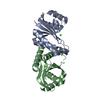
| ||||||||||||
|---|---|---|---|---|---|---|---|---|---|---|---|---|---|
| 1 |
| ||||||||||||
| Unit cell |
|
-Components
| #1: Protein | Mass: 19681.842 Da / Num. of mol.: 2 Source method: isolated from a genetically manipulated source Source: (gene. exp.) Deinococcus radiodurans (radioresistant)Strain: ATCC 13939 / DSM 20539 / JCM 16871 / LMG 4051 / NBRC 15346 / NCIMB 9279 / R1 / VKM B-1422 Gene: ruvC, DR_0440 / Production host: References: UniProt: Q9RX75, crossover junction endodeoxyribonuclease #2: Chemical | ChemComp-MG / |   Mass: 24.305 Da / Num. of mol.: 1 / Source method: isolated from a natural source / Formula: Mg / Feature type: SUBJECT OF INVESTIGATION Mass: 24.305 Da / Num. of mol.: 1 / Source method: isolated from a natural source / Formula: Mg / Feature type: SUBJECT OF INVESTIGATIONHas ligand of interest | Y | |
|---|
-Experimental details
-Experiment
| Experiment | Method: X-RAY DIFFRACTION / Number of used crystals: 1 |
|---|
- Sample preparation
| Crystal | Density Matthews: 2.14 Å3/Da / Density % sol: 42.48 % |
|---|---|
| Crystal grow | Temperature: 289 K / Method: vapor diffusion, sitting drop / pH: 7.8 / Details: 0.2 M MgCl2, 0.1M HEPES (pH 7.8), 20% PEG 3350 |
-Data collection
| Diffraction | Mean temperature: 80 K / Serial crystal experiment: N |
|---|---|
| Diffraction source | Source: SYNCHROTRON / Site: SSRF / Beamline: BL02U1 / Wavelength: 0.9792 Å |
| Detector | Type: ADSC QUANTUM 315r / Detector: CCD / Date: Nov 20, 2021 |
| Radiation | Protocol: SINGLE WAVELENGTH / Monochromatic (M) / Laue (L): M / Scattering type: x-ray |
| Radiation wavelength | Wavelength: 0.9792 Å / Relative weight: 1 |
| Reflection | Resolution: 2.75→27.71 Å / Num. obs: 9594 / % possible obs: 91.2 % / Redundancy: 4 % / Biso Wilson estimate: 57.4767212532 Å2 / Rmerge(I) obs: 0.131 / Net I/σ(I): 13.4 |
| Reflection shell | Resolution: 2.75→2.79 Å / Rmerge(I) obs: 0.722 / Num. unique obs: 674 |
- Processing
| Software |
| ||||||||||||||||||||||||||||||||||||||||
|---|---|---|---|---|---|---|---|---|---|---|---|---|---|---|---|---|---|---|---|---|---|---|---|---|---|---|---|---|---|---|---|---|---|---|---|---|---|---|---|---|---|
| Refinement | Method to determine structure: MOLECULAR REPLACEMENT Starting model: 4EP4 Resolution: 2.75016129042→27.705522236 Å / SU ML: 0.400409488542 / Cross valid method: FREE R-VALUE / σ(F): 1.3549347961 / Phase error: 32.9446404357 Stereochemistry target values: GeoStd + Monomer Library + CDL v1.2
| ||||||||||||||||||||||||||||||||||||||||
| Solvent computation | Shrinkage radii: 0.9 Å / VDW probe radii: 1.11 Å / Solvent model: FLAT BULK SOLVENT MODEL | ||||||||||||||||||||||||||||||||||||||||
| Displacement parameters | Biso mean: 63.2475739321 Å2 | ||||||||||||||||||||||||||||||||||||||||
| Refinement step | Cycle: LAST / Resolution: 2.75016129042→27.705522236 Å
| ||||||||||||||||||||||||||||||||||||||||
| Refine LS restraints |
| ||||||||||||||||||||||||||||||||||||||||
| LS refinement shell |
| ||||||||||||||||||||||||||||||||||||||||
| Refinement TLS params. | Method: refined / Origin x: 18.9841384343 Å / Origin y: 24.6401043917 Å / Origin z: 11.5409278192 Å
| ||||||||||||||||||||||||||||||||||||||||
| Refinement TLS group | Selection details: all |
