 Movie
Movie Controller


+ Open data

- Basic information
| Entry | Database: PDB / ID: 7w89 | ||||||
|---|---|---|---|---|---|---|---|
| Title | The structure of Deinococcus radiodurans Yqgf | ||||||
 Components Components | Putative pre-16S rRNA nuclease | ||||||
 Keywords Keywords | HYDROLASE / RNase / RNA binding | ||||||
| Function / homology |  Function and homology information Function and homology informationrRNA 5'-end processing / exonuclease activity / endonuclease activity / Hydrolases; Acting on ester bonds / cytoplasm Similarity search - Function | ||||||
| Biological species |  Deinococcus radiodurans (radioresistant) Deinococcus radiodurans (radioresistant) | ||||||
| Method |  X-RAY DIFFRACTION / SYNCHROTRON / MOLECULAR REPLACEMENT / Resolution: 1.50006105724 Å X-RAY DIFFRACTION / SYNCHROTRON / MOLECULAR REPLACEMENT / Resolution: 1.50006105724 Å | ||||||
 Authors Authors | Cheng, K. | ||||||
| Funding support |  China, 1items China, 1items
| ||||||
 Citation Citation | Journal: Mbio / Year: 2022 Title: Biochemical and Structural Study of RuvC and YqgF from Deinococcus radiodurans. Authors: Sun, Y. / Yang, J. / Xu, G. / Cheng, K. | ||||||
| History |
|
- Structure visualization
| Structure viewer | Molecule: MolmilJmol/JSmol |
|---|
- Downloads & links
-Download
| PDBx/mmCIF format | 7w89.cif.gz | 71.8 KB | Display | PDBx/mmCIF format |
|---|---|---|---|---|
| PDB format | pdb7w89.ent.gz | 45.7 KB | Display | PDB format |
| PDBx/mmJSON format | 7w89.json.gz | Tree view | PDBx/mmJSON format | |
| Others |  Other downloads Other downloads |
-Validation report
| Arichive directory | https://data.pdbj.org/pub/pdb/validation_reports/w8/7w89ftp://data.pdbj.org/pub/pdb/validation_reports/w8/7w89 | HTTPS FTP |
|---|
-Related structure data
| Related structure data | 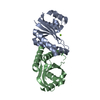 7w8dC 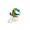 1ovqS S: Starting model for refinement C: citing same article ( |
|---|---|
| Similar structure data |
-Links
 PDBj
PDBj- Assembly
| Deposited unit | 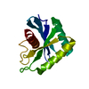
| ||||||||||||
|---|---|---|---|---|---|---|---|---|---|---|---|---|---|
| 1 |
| ||||||||||||
| Unit cell |
| ||||||||||||
| Components on special symmetry positions |
|
-Components
| #1: Protein | Mass: 14841.159 Da / Num. of mol.: 1 Source method: isolated from a genetically manipulated source Source: (gene. exp.) Deinococcus radiodurans (radioresistant)Strain: ATCC 13939 / DSM 20539 / JCM 16871 / LMG 4051 / NBRC 15346 / NCIMB 9279 / R1 / VKM B-1422 Gene: DR_2509 / Production host: References: UniProt: Q9RRI2, Hydrolases; Acting on ester bonds |
|---|---|
| #2: Water | ChemComp-HOH /  Mass: 18.015 Da / Num. of mol.: 156 / Source method: isolated from a natural source / Formula: H2O Mass: 18.015 Da / Num. of mol.: 156 / Source method: isolated from a natural source / Formula: H2O |
-Experimental details
-Experiment
| Experiment | Method: X-RAY DIFFRACTION / Number of used crystals: 1 |
|---|
- Sample preparation
| Crystal | Density Matthews: 2 Å3/Da / Density % sol: 38.35 % |
|---|---|
| Crystal grow | Temperature: 289 K / Method: vapor diffusion, sitting drop / pH: 7 / Details: 0.1 M MgCl2, 0.1M HEPES (pH 7.0), 15% PEG 4000 |
-Data collection
| Diffraction | Mean temperature: 80 K / Serial crystal experiment: N |
|---|---|
| Diffraction source | Source: SYNCHROTRON / Site: SSRF / Beamline: BL02U1 / Wavelength: 0.9792 Å |
| Detector | Type: ADSC QUANTUM 315r / Detector: CCD / Date: Nov 20, 2021 |
| Radiation | Protocol: SINGLE WAVELENGTH / Monochromatic (M) / Laue (L): M / Scattering type: x-ray |
| Radiation wavelength | Wavelength: 0.9792 Å / Relative weight: 1 |
| Reflection | Resolution: 1.5→27.07 Å / Num. obs: 19580 / % possible obs: 93.7 % / Redundancy: 4.5 % / Biso Wilson estimate: 16.5 Å2 / Rmerge(I) obs: 0.055 / Net I/σ(I): 26.04 |
| Reflection shell | Resolution: 1.5→1.54 Å / Rmerge(I) obs: 0.132 / Num. unique obs: 1430 |
- Processing
| Software |
| |||||||||||||||||||||||||||||||||||||||||||||||||
|---|---|---|---|---|---|---|---|---|---|---|---|---|---|---|---|---|---|---|---|---|---|---|---|---|---|---|---|---|---|---|---|---|---|---|---|---|---|---|---|---|---|---|---|---|---|---|---|---|---|---|
| Refinement | Method to determine structure: MOLECULAR REPLACEMENT Starting model: 1OVQ Resolution: 1.50006105724→27.0688711921 Å / SU ML: 0.11487136053 / Cross valid method: FREE R-VALUE / σ(F): 1.4025631195 / Phase error: 18.1688009337 Stereochemistry target values: GeoStd + Monomer Library + CDL v1.2
| |||||||||||||||||||||||||||||||||||||||||||||||||
| Solvent computation | Shrinkage radii: 0.9 Å / VDW probe radii: 1.11 Å / Solvent model: FLAT BULK SOLVENT MODEL | |||||||||||||||||||||||||||||||||||||||||||||||||
| Displacement parameters | Biso mean: 15.6326682596 Å2 | |||||||||||||||||||||||||||||||||||||||||||||||||
| Refinement step | Cycle: LAST / Resolution: 1.50006105724→27.0688711921 Å
| |||||||||||||||||||||||||||||||||||||||||||||||||
| Refine LS restraints |
| |||||||||||||||||||||||||||||||||||||||||||||||||
| LS refinement shell |
| |||||||||||||||||||||||||||||||||||||||||||||||||
| Refinement TLS params. | Method: refined / Origin x: 79.2987867651 Å / Origin y: 0.666036948163 Å / Origin z: 41.8795882323 Å
| |||||||||||||||||||||||||||||||||||||||||||||||||
| Refinement TLS group | Selection details: all |
