 Movie
Movie Controller

[English] 日本語
 Yorodumi
- PDB-7w71: Crystal structure of the PDZ-C domain of E. coli RseP in complex ... -
+ Open data

- Basic information
| Entry | Database: PDB / ID: 7w71 | |||||||||||||||
|---|---|---|---|---|---|---|---|---|---|---|---|---|---|---|---|---|
| Title | Crystal structure of the PDZ-C domain of E. coli RseP in complex with 12C7 Fab | |||||||||||||||
 Components Components |
| |||||||||||||||
 Keywords Keywords | HYDROLASE/IMMUNE SYSTEM / Intramembrane protease / HYDROLASE / HYDROLASE-IMMUNE SYSTEM complex | |||||||||||||||
| Function / homology |  Function and homology information Function and homology informationanti-sigma factor antagonist activity / Hydrolases; Acting on peptide bonds (peptidases); Metalloendopeptidases / cellular response to cell envelope stress / metalloendopeptidase activity / positive regulation of DNA-templated transcription / proteolysis / metal ion binding / plasma membrane Similarity search - Function | |||||||||||||||
| Biological species |   | |||||||||||||||
| Method |  X-RAY DIFFRACTION / SYNCHROTRON / MOLECULAR REPLACEMENT / Resolution: 3.2 Å X-RAY DIFFRACTION / SYNCHROTRON / MOLECULAR REPLACEMENT / Resolution: 3.2 Å | |||||||||||||||
 Authors Authors | Hirose, T. / Katagiri, S. / Nogi, T. | |||||||||||||||
| Funding support |  Japan, 4items Japan, 4items
| |||||||||||||||
 Citation Citation | Journal: Sci Adv / Year: 2022 Title: Mechanistic insights into intramembrane proteolysis by site-2 protease homolog RseP. Authors: Yuki Imaizumi / Kazunori Takanuki / Takuya Miyake / Mizuki Takemoto / Kunio Hirata / Mika Hirose / Rika Oi / Tatsuya Kobayashi / Kenichi Miyoshi / Rie Aruga / Tatsuhiko Yokoyama / Shizuka ...Authors: Yuki Imaizumi / Kazunori Takanuki / Takuya Miyake / Mizuki Takemoto / Kunio Hirata / Mika Hirose / Rika Oi / Tatsuya Kobayashi / Kenichi Miyoshi / Rie Aruga / Tatsuhiko Yokoyama / Shizuka Katagiri / Hiroaki Matsuura / Kenji Iwasaki / Takayuki Kato / Mika K Kaneko / Yukinari Kato / Michiko Tajiri / Satoko Akashi / Osamu Nureki / Yohei Hizukuri / Yoshinori Akiyama / Terukazu Nogi / Abstract: Site-2 proteases are a conserved family of intramembrane proteases that cleave transmembrane substrates to regulate signal transduction and maintain proteostasis. Here, we elucidated crystal ...Site-2 proteases are a conserved family of intramembrane proteases that cleave transmembrane substrates to regulate signal transduction and maintain proteostasis. Here, we elucidated crystal structures of inhibitor-bound forms of bacterial site-2 proteases including RseP. Structure-based chemical modification and cross-linking experiments indicated that the RseP domains surrounding the active center undergo conformational changes to expose the substrate-binding site, suggesting that RseP has a gating mechanism to regulate substrate entry. Furthermore, mutational analysis suggests that a conserved electrostatic linkage between the transmembrane and peripheral membrane-associated domains mediates the conformational changes. In vivo cleavage assays also support that the substrate transmembrane helix is unwound by strand addition to the intramembrane β sheet of RseP and is clamped by a conserved asparagine residue at the active center for efficient cleavage. This mechanism underlying the substrate binding, i.e., unwinding and clamping, appears common across distinct families of intramembrane proteases that cleave transmembrane segments. | |||||||||||||||
| History |
|
- Structure visualization
| Structure viewer | Molecule: MolmilJmol/JSmol |
|---|
- Downloads & links
-Download
| PDBx/mmCIF format | 7w71.cif.gz | 250.8 KB | Display | PDBx/mmCIF format |
|---|---|---|---|---|
| PDB format | pdb7w71.ent.gz | 162.9 KB | Display | PDB format |
| PDBx/mmJSON format | 7w71.json.gz | Tree view | PDBx/mmJSON format | |
| Others |  Other downloads Other downloads |
-Validation report
| Summary document | 7w71_validation.pdf.gz | 464.5 KB | Display | wwPDB validaton report |
|---|---|---|---|---|
| Full document | 7w71_full_validation.pdf.gz | 474.2 KB | Display | |
| Data in XML | 7w71_validation.xml.gz | 34.9 KB | Display | |
| Data in CIF | 7w71_validation.cif.gz | 47.4 KB | Display | |
| Arichive directory | https://data.pdbj.org/pub/pdb/validation_reports/w7/7w71ftp://data.pdbj.org/pub/pdb/validation_reports/w7/7w71 | HTTPS FTP |
-Related structure data
| Related structure data | 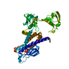 7w6xC  7w6yC  7w6zC  7w70C 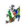 2zpmS  3wkmS S: Starting model for refinement C: citing same article ( |
|---|---|
| Similar structure data |


-Links
 PDBj
PDBj


- Assembly
| Deposited unit | 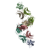
| ||||||||||||
|---|---|---|---|---|---|---|---|---|---|---|---|---|---|
| 1 | 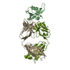
| ||||||||||||
| 2 | 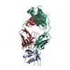
| ||||||||||||
| Unit cell |
|
-Components
| #1: Protein | Mass: 9765.188 Da / Num. of mol.: 2 Source method: isolated from a genetically manipulated source Source: (gene. exp.) References: UniProt: P0AEH1, Hydrolases; Acting on peptide bonds (peptidases); Metalloendopeptidases #2: Antibody | Mass: 23427.219 Da / Num. of mol.: 2 Source method: isolated from a genetically manipulated source Source: (gene. exp.) #3: Antibody | Mass: 23668.957 Da / Num. of mol.: 2 Source method: isolated from a genetically manipulated source Source: (gene. exp.) Has protein modification | Y | |
|---|
-Experimental details
-Experiment
| Experiment | Method: X-RAY DIFFRACTION / Number of used crystals: 1 |
|---|
- Sample preparation
| Crystal | Density Matthews: 2.8 Å3/Da / Density % sol: 56.06 % Description: The entry contains friedel pairs in F_plus/minus columns and I_plus/minus columns |
|---|---|
| Crystal grow | Temperature: 293 K / Method: vapor diffusion, hanging drop / pH: 5.5 Details: 25% (wt./vol.) Polyethylene glycol 10000, 0.2 M ammonium sulfate, 0.1 M Bis-Tris-HCl (pH 5.5) |
-Data collection
| Diffraction | Mean temperature: 100 K / Serial crystal experiment: N |
|---|---|
| Diffraction source | Source: SYNCHROTRON / Site: Photon Factory / Beamline: BL-17A / Wavelength: 0.98 Å |
| Detector | Type: DECTRIS PILATUS 6M / Detector: PIXEL / Date: Jun 25, 2019 |
| Radiation | Protocol: SINGLE WAVELENGTH / Monochromatic (M) / Laue (L): M / Scattering type: x-ray |
| Radiation wavelength | Wavelength: 0.98 Å / Relative weight: 1 |
| Reflection | Resolution: 3.2→49.5 Å / Num. obs: 21990 / % possible obs: 99.9 % / Redundancy: 6.8 % / Biso Wilson estimate: 64.8 Å2 / CC1/2: 0.992 / Rpim(I) all: 0.105 / Net I/σ(I): 7.5 |
| Reflection shell | Resolution: 3.2→3.42 Å / Mean I/σ(I) obs: 1.1 / Num. unique obs: 3909 / CC1/2: 0.738 / Rpim(I) all: 0.447 |
- Processing
| Software |
| ||||||||||||||||||||||||||||||||||||||||||||||||||||||||||||||||||||||||||||||||||||||||||||||||||||||||||||||||
|---|---|---|---|---|---|---|---|---|---|---|---|---|---|---|---|---|---|---|---|---|---|---|---|---|---|---|---|---|---|---|---|---|---|---|---|---|---|---|---|---|---|---|---|---|---|---|---|---|---|---|---|---|---|---|---|---|---|---|---|---|---|---|---|---|---|---|---|---|---|---|---|---|---|---|---|---|---|---|---|---|---|---|---|---|---|---|---|---|---|---|---|---|---|---|---|---|---|---|---|---|---|---|---|---|---|---|---|---|---|---|---|---|---|
| Refinement | Method to determine structure: MOLECULAR REPLACEMENT Starting model: 2ZPM, 3WKM Resolution: 3.2→49.5 Å / SU ML: 0.5509 / Cross valid method: FREE R-VALUE / σ(F): 0.11 / Phase error: 33.6419 Stereochemistry target values: GeoStd + Monomer Library + CDL v1.2 Details: The entry contains friedel pairs in F_plus/minus columns and I_plus/minus columns
| ||||||||||||||||||||||||||||||||||||||||||||||||||||||||||||||||||||||||||||||||||||||||||||||||||||||||||||||||
| Solvent computation | Shrinkage radii: 0.9 Å / VDW probe radii: 1.11 Å / Solvent model: FLAT BULK SOLVENT MODEL | ||||||||||||||||||||||||||||||||||||||||||||||||||||||||||||||||||||||||||||||||||||||||||||||||||||||||||||||||
| Displacement parameters | Biso mean: 65.06 Å2 | ||||||||||||||||||||||||||||||||||||||||||||||||||||||||||||||||||||||||||||||||||||||||||||||||||||||||||||||||
| Refinement step | Cycle: LAST / Resolution: 3.2→49.5 Å
| ||||||||||||||||||||||||||||||||||||||||||||||||||||||||||||||||||||||||||||||||||||||||||||||||||||||||||||||||
| Refine LS restraints |
| ||||||||||||||||||||||||||||||||||||||||||||||||||||||||||||||||||||||||||||||||||||||||||||||||||||||||||||||||
| LS refinement shell |
|
