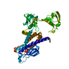
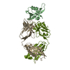
Journal: Sci Adv / Year: 2022 Title: Mechanistic insights into intramembrane proteolysis by site-2 protease homolog RseP. Authors: Yuki Imaizumi / Kazunori Takanuki / Takuya Miyake / Mizuki Takemoto / Kunio Hirata / Mika Hirose / Rika Oi / Tatsuya Kobayashi / Kenichi Miyoshi / Rie Aruga / Tatsuhiko Yokoyama / Shizuka ...Authors: Yuki Imaizumi / Kazunori Takanuki / Takuya Miyake / Mizuki Takemoto / Kunio Hirata / Mika Hirose / Rika Oi / Tatsuya Kobayashi / Kenichi Miyoshi / Rie Aruga / Tatsuhiko Yokoyama / Shizuka Katagiri / Hiroaki Matsuura / Kenji Iwasaki / Takayuki Kato / Mika K Kaneko / Yukinari Kato / Michiko Tajiri / Satoko Akashi / Osamu Nureki / Yohei Hizukuri / Yoshinori Akiyama / Terukazu Nogi / Abstract: Site-2 proteases are a conserved family of intramembrane proteases that cleave transmembrane substrates to regulate signal transduction and maintain proteostasis. Here, we elucidated crystal ...Site-2 proteases are a conserved family of intramembrane proteases that cleave transmembrane substrates to regulate signal transduction and maintain proteostasis. Here, we elucidated crystal structures of inhibitor-bound forms of bacterial site-2 proteases including RseP. Structure-based chemical modification and cross-linking experiments indicated that the RseP domains surrounding the active center undergo conformational changes to expose the substrate-binding site, suggesting that RseP has a gating mechanism to regulate substrate entry. Furthermore, mutational analysis suggests that a conserved electrostatic linkage between the transmembrane and peripheral membrane-associated domains mediates the conformational changes. In vivo cleavage assays also support that the substrate transmembrane helix is unwound by strand addition to the intramembrane β sheet of RseP and is clamped by a conserved asparagine residue at the active center for efficient cleavage. This mechanism underlying the substrate binding, i.e., unwinding and clamping, appears common across distinct families of intramembrane proteases that cleave transmembrane segments.
In the structure databanks used in Yorodumi, some data are registered as the other names, "COVID-19 virus" and "2019-nCoV". Here are the details of the virus and the list of structure data.
Jan 31, 2019. EMDB accession codes are about to change! (news from PDBe EMDB page)
EMDB accession codes are about to change! (news from PDBe EMDB page)
The allocation of 4 digits for EMDB accession codes will soon come to an end. Whilst these codes will remain in use, new EMDB accession codes will include an additional digit and will expand incrementally as the available range of codes is exhausted. The current 4-digit format prefixed with “EMD-” (i.e. EMD-XXXX) will advance to a 5-digit format (i.e. EMD-XXXXX), and so on. It is currently estimated that the 4-digit codes will be depleted around Spring 2019, at which point the 5-digit format will come into force.
The EM Navigator/Yorodumi systems omit the EMD- prefix.
Related info.:Q: What is EMD? / ID/Accession-code notation in Yorodumi/EM Navigator
Yorodumi is a browser for structure data from EMDB, PDB, SASBDB, etc.
This page is also the successor to EM Navigator detail page, and also detail information page/front-end page for Omokage search.
The word "yorodu" (or yorozu) is an old Japanese word meaning "ten thousand". "mi" (miru) is to see.
Related info.:EMDB / PDB / SASBDB / Comparison of 3 databanks / Yorodumi Search / Aug 31, 2016. New EM Navigator & Yorodumi / Yorodumi Papers / Jmol/JSmol / Function and homology information / Changes in new EM Navigator and Yorodumi
 Movie
Movie Controller

 Yorodumi
 Open data
 Basic information
 Components
 Keywords
 Function and homology information
 Kangiella koreensis DSM 16069 (bacteria)
 X-RAY DIFFRACTION /
  Authors
 Japan, 4items
  Citation
 Structure visualization
 Downloads & links
 Other downloads


 PDBj
PDBj
 Assembly

 Mass: 65.409 Da / Num. of mol.: 1 / Source method: obtained synthetically / Formula: Zn
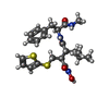
 Mass: 477.640 Da / Num. of mol.: 1 / Source method: obtained synthetically / Formula: C23H31N3O4S2 / Comment: inhibitor*YM
 Sample preparation
 Processing