 Movie
Movie Controller

[English] 日本語
 Yorodumi
- PDB-7w28: Crystal Structure of SETD3-SAH in complex with betaA-4PyrAla73 peptide -
+ Open data

- Basic information
| Entry | Database: PDB / ID: 7w28 | ||||||
|---|---|---|---|---|---|---|---|
| Title | Crystal Structure of SETD3-SAH in complex with betaA-4PyrAla73 peptide | ||||||
 Components Components |
| ||||||
 Keywords Keywords | STRUCTURAL PROTEIN / SET domain | ||||||
| Function / homology |  Function and homology information Function and homology informationpeptidyl-histidine methylation / regulation of uterine smooth muscle contraction / protein-histidine N-methyltransferase / protein-L-histidine N-tele-methyltransferase activity / actin modification / positive regulation of norepinephrine uptake / bBAF complex / cellular response to cytochalasin B / npBAF complex / nBAF complex ...peptidyl-histidine methylation / regulation of uterine smooth muscle contraction / protein-histidine N-methyltransferase / protein-L-histidine N-tele-methyltransferase activity / actin modification / positive regulation of norepinephrine uptake / bBAF complex / cellular response to cytochalasin B / npBAF complex / nBAF complex / brahma complex / histone H3K36 methyltransferase activity / regulation of transepithelial transport / Formation of annular gap junctions / Formation of the dystrophin-glycoprotein complex (DGC) / morphogenesis of a polarized epithelium / structural constituent of postsynaptic actin cytoskeleton / Gap junction degradation / GBAF complex / Folding of actin by CCT/TriC / regulation of G0 to G1 transition / protein localization to adherens junction / Cell-extracellular matrix interactions / dense body / Tat protein binding / postsynaptic actin cytoskeleton / Prefoldin mediated transfer of substrate to CCT/TriC / RSC-type complex / regulation of double-strand break repair / regulation of nucleotide-excision repair / histone H3K4 methyltransferase activity / Adherens junctions interactions / RHOF GTPase cycle / adherens junction assembly / apical protein localization / Sensory processing of sound by outer hair cells of the cochlea / Interaction between L1 and Ankyrins / tight junction / SWI/SNF complex / regulation of mitotic metaphase/anaphase transition / Sensory processing of sound by inner hair cells of the cochlea / positive regulation of T cell differentiation / positive regulation of muscle cell differentiation / apical junction complex / positive regulation of double-strand break repair / maintenance of blood-brain barrier / regulation of norepinephrine uptake / nitric-oxide synthase binding / transporter regulator activity / cortical cytoskeleton / NuA4 histone acetyltransferase complex / establishment or maintenance of cell polarity / positive regulation of stem cell population maintenance / Recycling pathway of L1 / Regulation of MITF-M-dependent genes involved in pigmentation / brush border / regulation of G1/S transition of mitotic cell cycle / EPH-ephrin mediated repulsion of cells / negative regulation of cell differentiation / kinesin binding / RHO GTPases Activate WASPs and WAVEs / regulation of synaptic vesicle endocytosis / positive regulation of myoblast differentiation / RHO GTPases activate IQGAPs / regulation of protein localization to plasma membrane / positive regulation of double-strand break repair via homologous recombination / EPHB-mediated forward signaling / cytoskeleton organization / substantia nigra development / axonogenesis / calyx of Held / nitric-oxide synthase regulator activity / Translocation of SLC2A4 (GLUT4) to the plasma membrane / FCGR3A-mediated phagocytosis / actin filament / adherens junction / Regulation of endogenous retroelements by Piwi-interacting RNAs (piRNAs) / positive regulation of cell differentiation / cell motility / RHO GTPases Activate Formins / Signaling by high-kinase activity BRAF mutants / MAP2K and MAPK activation / DNA Damage Recognition in GG-NER / Regulation of actin dynamics for phagocytic cup formation / B-WICH complex positively regulates rRNA expression / kinetochore / PKMTs methylate histone lysines / structural constituent of cytoskeleton / Hydrolases; Acting on acid anhydrides; Acting on acid anhydrides to facilitate cellular and subcellular movement / VEGFA-VEGFR2 Pathway / platelet aggregation / Schaffer collateral - CA1 synapse / tau protein binding / nuclear matrix / cytoplasmic ribonucleoprotein granule / Signaling by RAF1 mutants / Signaling by moderate kinase activity BRAF mutants / Paradoxical activation of RAF signaling by kinase inactive BRAF / Signaling downstream of RAS mutants / cell-cell junction Similarity search - Function | ||||||
| Biological species |  Homo sapiens (human) Homo sapiens (human) | ||||||
| Method |  X-RAY DIFFRACTION / SYNCHROTRON / MOLECULAR REPLACEMENT / Resolution: 1.79 Å X-RAY DIFFRACTION / SYNCHROTRON / MOLECULAR REPLACEMENT / Resolution: 1.79 Å | ||||||
 Authors Authors | Li, H. / Ma, H. | ||||||
| Funding support |  China, 1items China, 1items
| ||||||
 Citation Citation | Journal: Protein Sci. / Year: 2022 Title: Histidine methyltransferase SETD3 methylates structurally diverse histidine mimics in actin. Authors: Hintzen, J.C.J. / Ma, H. / Deng, H. / Witecka, A. / Andersen, S.B. / Drozak, J. / Guo, H. / Qian, P. / Li, H. / Mecinovic, J. | ||||||
| History |
|
- Structure visualization
| Structure viewer | Molecule: MolmilJmol/JSmol |
|---|
- Downloads & links
-Download
| PDBx/mmCIF format | 7w28.cif.gz | 232.3 KB | Display | PDBx/mmCIF format |
|---|---|---|---|---|
| PDB format | pdb7w28.ent.gz | 181.6 KB | Display | PDB format |
| PDBx/mmJSON format | 7w28.json.gz | Tree view | PDBx/mmJSON format | |
| Others |  Other downloads Other downloads |
-Validation report
| Summary document | 7w28_validation.pdf.gz | 812.8 KB | Display | wwPDB validaton report |
|---|---|---|---|---|
| Full document | 7w28_full_validation.pdf.gz | 813.7 KB | Display | |
| Data in XML | 7w28_validation.xml.gz | 24.4 KB | Display | |
| Data in CIF | 7w28_validation.cif.gz | 37.3 KB | Display | |
| Arichive directory | https://data.pdbj.org/pub/pdb/validation_reports/w2/7w28ftp://data.pdbj.org/pub/pdb/validation_reports/w2/7w28 | HTTPS FTP |
-Related structure data
| Related structure data | 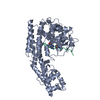 7w29C  6mbjS S: Starting model for refinement C: citing same article ( |
|---|---|
| Similar structure data |
-Links
 PDBj
PDBj


- Assembly
| Deposited unit | 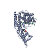
| ||||||||
|---|---|---|---|---|---|---|---|---|---|
| 1 |
| ||||||||
| Unit cell |
|
-Components
| #1: Protein | Mass: 57223.000 Da / Num. of mol.: 1 / Fragment: SETD3 Source method: isolated from a genetically manipulated source Source: (gene. exp.) Homo sapiens (human) / Gene: SETD3 / Plasmid: pSUMOH10 / Production host:  References: UniProt: Q86TU7, histone-lysine N-methyltransferase | ||||
|---|---|---|---|---|---|
| #2: Protein/peptide | Mass: 1913.111 Da / Num. of mol.: 1 / Fragment: SAH / Source method: obtained synthetically / Source: (synth.) Homo sapiens (human) / References: UniProt: P60709 | ||||
| #3: Chemical | ChemComp-SAH /   Type: L-peptide linking / Mass: 384.411 Da / Num. of mol.: 1 / Source method: obtained synthetically / Formula: C14H20N6O5S / Feature type: SUBJECT OF INVESTIGATION Type: L-peptide linking / Mass: 384.411 Da / Num. of mol.: 1 / Source method: obtained synthetically / Formula: C14H20N6O5S / Feature type: SUBJECT OF INVESTIGATION | ||||
| #4: Chemical |   Mass: 96.063 Da / Num. of mol.: 2 / Source method: obtained synthetically / Formula: SO4 Mass: 96.063 Da / Num. of mol.: 2 / Source method: obtained synthetically / Formula: SO4#5: Water | ChemComp-HOH / |  Mass: 18.015 Da / Num. of mol.: 514 / Source method: isolated from a natural source / Formula: H2O Mass: 18.015 Da / Num. of mol.: 514 / Source method: isolated from a natural source / Formula: H2OHas ligand of interest | Y | |
-Experimental details
-Experiment
| Experiment | Method: X-RAY DIFFRACTION / Number of used crystals: 1 |
|---|
- Sample preparation
| Crystal | Density Matthews: 2.25 Å3/Da / Density % sol: 45.26 % |
|---|---|
| Crystal grow | Temperature: 289 K / Method: vapor diffusion, sitting drop / pH: 8.5 Details: 30%(w/v) PEG4000, 0.1 M Tris base/Hydrochloric acid 8.5, 0.2M Lithium sulfate. |
-Data collection
| Diffraction | Mean temperature: 100 K / Serial crystal experiment: N | |||||||||||||||||||||||||||||||||||||||||||||||||||||||||||||||||||||||||||||||||||||||||||||||||||||||||||||||||||||||||||||||||||||||||||||||||||||||||||||||||||||||||||||||||||||||||||||
|---|---|---|---|---|---|---|---|---|---|---|---|---|---|---|---|---|---|---|---|---|---|---|---|---|---|---|---|---|---|---|---|---|---|---|---|---|---|---|---|---|---|---|---|---|---|---|---|---|---|---|---|---|---|---|---|---|---|---|---|---|---|---|---|---|---|---|---|---|---|---|---|---|---|---|---|---|---|---|---|---|---|---|---|---|---|---|---|---|---|---|---|---|---|---|---|---|---|---|---|---|---|---|---|---|---|---|---|---|---|---|---|---|---|---|---|---|---|---|---|---|---|---|---|---|---|---|---|---|---|---|---|---|---|---|---|---|---|---|---|---|---|---|---|---|---|---|---|---|---|---|---|---|---|---|---|---|---|---|---|---|---|---|---|---|---|---|---|---|---|---|---|---|---|---|---|---|---|---|---|---|---|---|---|---|---|---|---|---|---|---|
| Diffraction source | Source: SYNCHROTRON / Site: SSRF / Beamline: BL19U1 / Wavelength: 0.9788 Å | |||||||||||||||||||||||||||||||||||||||||||||||||||||||||||||||||||||||||||||||||||||||||||||||||||||||||||||||||||||||||||||||||||||||||||||||||||||||||||||||||||||||||||||||||||||||||||||
| Detector | Type: DECTRIS PILATUS3 X 6M / Detector: PIXEL / Date: Oct 22, 2020 / Details: mirrors | |||||||||||||||||||||||||||||||||||||||||||||||||||||||||||||||||||||||||||||||||||||||||||||||||||||||||||||||||||||||||||||||||||||||||||||||||||||||||||||||||||||||||||||||||||||||||||||
| Radiation | Protocol: SINGLE WAVELENGTH / Monochromatic (M) / Laue (L): M / Scattering type: x-ray | |||||||||||||||||||||||||||||||||||||||||||||||||||||||||||||||||||||||||||||||||||||||||||||||||||||||||||||||||||||||||||||||||||||||||||||||||||||||||||||||||||||||||||||||||||||||||||||
| Radiation wavelength | Wavelength: 0.9788 Å / Relative weight: 1 | |||||||||||||||||||||||||||||||||||||||||||||||||||||||||||||||||||||||||||||||||||||||||||||||||||||||||||||||||||||||||||||||||||||||||||||||||||||||||||||||||||||||||||||||||||||||||||||
| Reflection | Resolution: 1.79→50 Å / Num. obs: 49400 / % possible obs: 99.1 % / Redundancy: 5.1 % / Rmerge(I) obs: 0.122 / Rpim(I) all: 0.062 / Rrim(I) all: 0.138 / Χ2: 1.805 / Net I/σ(I): 6 | |||||||||||||||||||||||||||||||||||||||||||||||||||||||||||||||||||||||||||||||||||||||||||||||||||||||||||||||||||||||||||||||||||||||||||||||||||||||||||||||||||||||||||||||||||||||||||||
| Reflection shell | Diffraction-ID: 1
|
- Processing
| Software |
| ||||||||||||||||||||||||||||||||||||||||||||||||||||||||||||||||||||||||||||||||||||||||||||||||||||||||||||||||||||||||||||||
|---|---|---|---|---|---|---|---|---|---|---|---|---|---|---|---|---|---|---|---|---|---|---|---|---|---|---|---|---|---|---|---|---|---|---|---|---|---|---|---|---|---|---|---|---|---|---|---|---|---|---|---|---|---|---|---|---|---|---|---|---|---|---|---|---|---|---|---|---|---|---|---|---|---|---|---|---|---|---|---|---|---|---|---|---|---|---|---|---|---|---|---|---|---|---|---|---|---|---|---|---|---|---|---|---|---|---|---|---|---|---|---|---|---|---|---|---|---|---|---|---|---|---|---|---|---|---|---|
| Refinement | Method to determine structure: MOLECULAR REPLACEMENT Starting model: 6MBJ Resolution: 1.79→38.59 Å / SU ML: 0.21 / Cross valid method: THROUGHOUT / σ(F): 1.39 / Phase error: 20.81 / Stereochemistry target values: ML
| ||||||||||||||||||||||||||||||||||||||||||||||||||||||||||||||||||||||||||||||||||||||||||||||||||||||||||||||||||||||||||||||
| Solvent computation | Shrinkage radii: 0.9 Å / VDW probe radii: 1.11 Å / Solvent model: FLAT BULK SOLVENT MODEL | ||||||||||||||||||||||||||||||||||||||||||||||||||||||||||||||||||||||||||||||||||||||||||||||||||||||||||||||||||||||||||||||
| Displacement parameters | Biso max: 119.04 Å2 / Biso mean: 36.7786 Å2 / Biso min: 14.09 Å2 | ||||||||||||||||||||||||||||||||||||||||||||||||||||||||||||||||||||||||||||||||||||||||||||||||||||||||||||||||||||||||||||||
| Refinement step | Cycle: final / Resolution: 1.79→38.59 Å
| ||||||||||||||||||||||||||||||||||||||||||||||||||||||||||||||||||||||||||||||||||||||||||||||||||||||||||||||||||||||||||||||
| LS refinement shell | Refine-ID: X-RAY DIFFRACTION / Rfactor Rfree error: 0 / Total num. of bins used: 17
| ||||||||||||||||||||||||||||||||||||||||||||||||||||||||||||||||||||||||||||||||||||||||||||||||||||||||||||||||||||||||||||||
| Refinement TLS params. | Method: refined / Refine-ID: X-RAY DIFFRACTION
| ||||||||||||||||||||||||||||||||||||||||||||||||||||||||||||||||||||||||||||||||||||||||||||||||||||||||||||||||||||||||||||||
| Refinement TLS group |
|
