 Movie
Movie Controller

[English] 日本語
 Yorodumi
- PDB-7w1j: Crystal structure of carboxylesterase from Thermobifida fusca with J1K -
+ Open data

- Basic information
| Entry | Database: PDB / ID: 7w1j | ||||||
|---|---|---|---|---|---|---|---|
| Title | Crystal structure of carboxylesterase from Thermobifida fusca with J1K | ||||||
 Components Components | Carboxylesterase | ||||||
 Keywords Keywords | HYDROLASE / plastic degradation / catalysis | ||||||
| Function / homology |  Function and homology information Function and homology information | ||||||
| Biological species |   Thermobifida fusca (bacteria) Thermobifida fusca (bacteria) | ||||||
| Method |  X-RAY DIFFRACTION / SYNCHROTRON / MOLECULAR REPLACEMENT / Resolution: 1.92 Å X-RAY DIFFRACTION / SYNCHROTRON / MOLECULAR REPLACEMENT / Resolution: 1.92 Å | ||||||
 Authors Authors | Han, X. / Gerlis, H. / Li, Z. / Gao, J. / Wei, R. / Liu, W. | ||||||
| Funding support | 1items
| ||||||
 Citation Citation | Journal: Acs Catalysis / Year: 2022 Title: Structural Insights into (Tere)phthalate-Ester Hydrolysis by a Carboxylesterase and Its Role in Promoting PET Depolymerization Authors: Von Haugwitz, G. / Han, X. / Pfaff, L. / Li, Q. / Wei, H. / Gao, J. / Methling, K. / Ao, Y. / Brack, Y. / Mican, J. / Feiler, C.G. / Weiss, M.S. / Bednar, D. / Palm, G.J. / Lalk, M. / ...Authors: Von Haugwitz, G. / Han, X. / Pfaff, L. / Li, Q. / Wei, H. / Gao, J. / Methling, K. / Ao, Y. / Brack, Y. / Mican, J. / Feiler, C.G. / Weiss, M.S. / Bednar, D. / Palm, G.J. / Lalk, M. / Lammers, M. / Damborsky, J. / Weber, G. / Liu, W. / Bornscheuer, U.T. / Wei, R. | ||||||
| History |
|
- Structure visualization
| Structure viewer | Molecule: MolmilJmol/JSmol |
|---|
- Downloads & links
-Download
| PDBx/mmCIF format | 7w1j.cif.gz | 118.2 KB | Display | PDBx/mmCIF format |
|---|---|---|---|---|
| PDB format | pdb7w1j.ent.gz | 87 KB | Display | PDB format |
| PDBx/mmJSON format | 7w1j.json.gz | Tree view | PDBx/mmJSON format | |
| Others |  Other downloads Other downloads |
-Validation report
| Arichive directory | https://data.pdbj.org/pub/pdb/validation_reports/w1/7w1jftp://data.pdbj.org/pub/pdb/validation_reports/w1/7w1j | HTTPS FTP |
|---|
-Related structure data
| Related structure data | 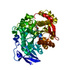 7w1iC 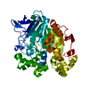 7w1kC 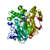 7w1lC 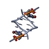 2gotS S: Starting model for refinement C: citing same article ( |
|---|---|
| Similar structure data |
-Links
 PDBj
PDBj
- Assembly
| Deposited unit | 
| ||||||||
|---|---|---|---|---|---|---|---|---|---|
| 1 |
| ||||||||
| Unit cell |
|
-Components
| #1: Protein | Mass: 52986.500 Da / Num. of mol.: 1 Source method: isolated from a genetically manipulated source Source: (gene. exp.) Thermobifida fusca (bacteria) / Production host: |
|---|---|
| #2: Chemical | ChemComp-J1K / 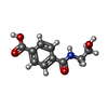  Mass: 209.199 Da / Num. of mol.: 1 / Source method: obtained synthetically / Formula: C10H11NO4 / Feature type: SUBJECT OF INVESTIGATION Mass: 209.199 Da / Num. of mol.: 1 / Source method: obtained synthetically / Formula: C10H11NO4 / Feature type: SUBJECT OF INVESTIGATION |
| #3: Water | ChemComp-HOH /  Mass: 18.015 Da / Num. of mol.: 444 / Source method: isolated from a natural source / Formula: H2O Mass: 18.015 Da / Num. of mol.: 444 / Source method: isolated from a natural source / Formula: H2O |
| Has ligand of interest | Y |
-Experimental details
-Experiment
| Experiment | Method: X-RAY DIFFRACTION / Number of used crystals: 1 |
|---|
- Sample preparation
| Crystal | Density Matthews: 2.36 Å3/Da / Density % sol: 47.95 % |
|---|---|
| Crystal grow | Temperature: 295 K / Method: vapor diffusion, sitting drop / pH: 7.5 Details: 25% w/v Poly(acrylic acid sodium salt) 5100, 0.02 M MgCl2, 0.1 M HEPES pH 7.5 |
-Data collection
| Diffraction | Mean temperature: 100 K / Serial crystal experiment: N |
|---|---|
| Diffraction source | Source: SYNCHROTRON / Site: BESSY  / Beamline: 14.1 / Wavelength: 1 Å / Beamline: 14.1 / Wavelength: 1 Å |
| Detector | Type: DECTRIS PILATUS3 2M / Detector: PIXEL / Date: Nov 15, 2020 |
| Radiation | Protocol: SINGLE WAVELENGTH / Monochromatic (M) / Laue (L): M / Scattering type: x-ray |
| Radiation wavelength | Wavelength: 1 Å / Relative weight: 1 |
| Reflection | Resolution: 1.92→30 Å / Num. obs: 38958 / % possible obs: 99.3 % / Redundancy: 13.2 % / CC1/2: 0.998 / Net I/σ(I): 15.1 |
| Reflection shell | Resolution: 1.92→2.03 Å / Num. unique obs: 5964 / CC1/2: 0.825 |
- Processing
| Software |
| ||||||||||||||||||||||||||||||||||||||||||||||||||||||||||||||||||
|---|---|---|---|---|---|---|---|---|---|---|---|---|---|---|---|---|---|---|---|---|---|---|---|---|---|---|---|---|---|---|---|---|---|---|---|---|---|---|---|---|---|---|---|---|---|---|---|---|---|---|---|---|---|---|---|---|---|---|---|---|---|---|---|---|---|---|---|
| Refinement | Method to determine structure: MOLECULAR REPLACEMENT Starting model: 2GOT Resolution: 1.92→30 Å / SU ML: 0.23 / Cross valid method: THROUGHOUT / σ(F): 1.35 / Phase error: 23.38 / Stereochemistry target values: ML
| ||||||||||||||||||||||||||||||||||||||||||||||||||||||||||||||||||
| Solvent computation | Shrinkage radii: 0.9 Å / VDW probe radii: 1.11 Å / Solvent model: FLAT BULK SOLVENT MODEL | ||||||||||||||||||||||||||||||||||||||||||||||||||||||||||||||||||
| Displacement parameters | Biso max: 85.68 Å2 / Biso mean: 31.0692 Å2 / Biso min: 16.93 Å2 | ||||||||||||||||||||||||||||||||||||||||||||||||||||||||||||||||||
| Refinement step | Cycle: final / Resolution: 1.92→30 Å
| ||||||||||||||||||||||||||||||||||||||||||||||||||||||||||||||||||
| LS refinement shell | Refine-ID: X-RAY DIFFRACTION / Rfactor Rfree error: 0
|
