 Movie
Movie Controller

[English] 日本語
 Yorodumi
- PDB-7vzf: Cryo-EM structure of amyloid fibril formed by full-length human SOD1 -
+ Open data

- Basic information
| Entry | Database: PDB / ID: 7vzf | ||||||||||||
|---|---|---|---|---|---|---|---|---|---|---|---|---|---|
| Title | Cryo-EM structure of amyloid fibril formed by full-length human SOD1 | ||||||||||||
 Components Components | Superoxide dismutase [Cu-Zn] | ||||||||||||
 Keywords Keywords | PROTEIN FIBRIL / Amyloid fibril | ||||||||||||
| Function / homology |  Function and homology information Function and homology informationaction potential initiation / response to antipsychotic drug / neurofilament cytoskeleton organization / response to carbon monoxide / relaxation of vascular associated smooth muscle / regulation of organ growth / dense core granule / response to superoxide / positive regulation of oxidative stress-induced intrinsic apoptotic signaling pathway / peripheral nervous system myelin maintenance ...action potential initiation / response to antipsychotic drug / neurofilament cytoskeleton organization / response to carbon monoxide / relaxation of vascular associated smooth muscle / regulation of organ growth / dense core granule / response to superoxide / positive regulation of oxidative stress-induced intrinsic apoptotic signaling pathway / peripheral nervous system myelin maintenance / anterograde axonal transport / protein phosphatase 2B binding / regulation of T cell differentiation in thymus / Oxidoreductases; Acting on a sulfur group of donors / regulation of GTPase activity / retina homeostasis / auditory receptor cell stereocilium organization / cellular response to potassium ion / hydrogen peroxide biosynthetic process / retrograde axonal transport / myeloid cell homeostasis / superoxide anion generation / superoxide metabolic process / muscle cell cellular homeostasis / response to copper ion / superoxide dismutase / Detoxification of Reactive Oxygen Species / superoxide dismutase activity / heart contraction / cellular response to cadmium ion / cellular response to ATP / negative regulation of reproductive process / negative regulation of developmental process / transmission of nerve impulse / regulation of multicellular organism growth / ectopic germ cell programmed cell death / response to axon injury / ovarian follicle development / neuronal action potential / positive regulation of superoxide anion generation / axon cytoplasm / removal of superoxide radicals / embryo implantation / reactive oxygen species metabolic process / Gene and protein expression by JAK-STAT signaling after Interleukin-12 stimulation / positive regulation of phagocytosis / dendrite cytoplasm / placenta development / thymus development / positive regulation of cytokine production / determination of adult lifespan / response to amphetamine / regulation of mitochondrial membrane potential / glutathione metabolic process / response to hydrogen peroxide / locomotory behavior / sensory perception of sound / response to nutrient levels / mitochondrial intermembrane space / negative regulation of inflammatory response / regulation of blood pressure / small GTPase binding / Platelet degranulation / peroxisome / response to heat / protein-folding chaperone binding / cytoplasmic vesicle / spermatogenesis / gene expression / intracellular iron ion homeostasis / response to ethanol / negative regulation of neuron apoptotic process / positive regulation of MAPK cascade / lysosome / positive regulation of apoptotic process / mitochondrial matrix / response to xenobiotic stimulus / copper ion binding / neuronal cell body / apoptotic process / protein homodimerization activity / protein-containing complex / mitochondrion / : / extracellular exosome / extracellular region / zinc ion binding / nucleoplasm / identical protein binding / nucleus / cytoplasm / cytosol Similarity search - Function | ||||||||||||
| Biological species |  Homo sapiens (human) Homo sapiens (human) | ||||||||||||
| Method | ELECTRON MICROSCOPY / helical reconstruction / cryo EM / Resolution: 2.95 Å | ||||||||||||
 Authors Authors | Wang, L.Q. / Ma, Y.Y. / Yuan, H.Y. / Zhao, K. / Zhang, M.Y. / Wang, Q. / Huang, X. / Xu, W.C. / Chen, J. / Li, D. ...Wang, L.Q. / Ma, Y.Y. / Yuan, H.Y. / Zhao, K. / Zhang, M.Y. / Wang, Q. / Huang, X. / Xu, W.C. / Chen, J. / Li, D. / Zhang, D.L. / Zou, L.Y. / Yin, P. / Liu, C. / Liang, Y. | ||||||||||||
| Funding support |  China, 3items China, 3items
| ||||||||||||
 Citation Citation | Journal: Nat Commun / Year: 2022 Title: Cryo-EM structure of an amyloid fibril formed by full-length human SOD1 reveals its conformational conversion. Authors: Li-Qiang Wang / Yeyang Ma / Han-Ye Yuan / Kun Zhao / Mu-Ya Zhang / Qiang Wang / Xi Huang / Wen-Chang Xu / Bin Dai / Jie Chen / Dan Li / Delin Zhang / Zhengzhi Wang / Liangyu Zou / Ping Yin / ...Authors: Li-Qiang Wang / Yeyang Ma / Han-Ye Yuan / Kun Zhao / Mu-Ya Zhang / Qiang Wang / Xi Huang / Wen-Chang Xu / Bin Dai / Jie Chen / Dan Li / Delin Zhang / Zhengzhi Wang / Liangyu Zou / Ping Yin / Cong Liu / Yi Liang / Abstract: Amyotrophic lateral sclerosis (ALS) is a neurodegenerative disease. Misfolded Cu, Zn-superoxide dismutase (SOD1) has been linked to both familial and sporadic ALS. SOD1 fibrils formed in vitro share ...Amyotrophic lateral sclerosis (ALS) is a neurodegenerative disease. Misfolded Cu, Zn-superoxide dismutase (SOD1) has been linked to both familial and sporadic ALS. SOD1 fibrils formed in vitro share toxic properties with ALS inclusions. Here we produced cytotoxic amyloid fibrils from full-length apo human SOD1 under reducing conditions and determined the atomic structure using cryo-EM. The SOD1 fibril consists of a single protofilament with a left-handed helix. The fibril core exhibits a serpentine fold comprising N-terminal segment (residues 3-55) and C-terminal segment (residues 86-153) with an intrinsic disordered segment. The two segments are zipped up by three salt bridge pairs. By comparison with the structure of apo SOD1 dimer, we propose that eight β-strands (to form a β-barrel) and one α-helix in the subunit of apo SOD1 convert into thirteen β-strands stabilized by five hydrophobic cavities in the SOD1 fibril. Our data provide insights into how SOD1 converts between structurally and functionally distinct states. | ||||||||||||
| History |
|
- Structure visualization
| Structure viewer | Molecule: MolmilJmol/JSmol |
|---|
- Downloads & links
-Download
| PDBx/mmCIF format | 7vzf.cif.gz | 68.7 KB | Display | PDBx/mmCIF format |
|---|---|---|---|---|
| PDB format | pdb7vzf.ent.gz | 51 KB | Display | PDB format |
| PDBx/mmJSON format | 7vzf.json.gz | Tree view | PDBx/mmJSON format | |
| Others |  Other downloads Other downloads |
-Validation report
| Arichive directory | https://data.pdbj.org/pub/pdb/validation_reports/vz/7vzfftp://data.pdbj.org/pub/pdb/validation_reports/vz/7vzf | HTTPS FTP |
|---|
-Related structure data
| Related structure data | 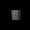 32227MC M: map data used to model this data C: citing same article ( |
|---|---|
| Similar structure data |
-Links
 PDBj
PDBj


- Assembly
| Deposited unit | 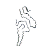
|
|---|---|
| 1 |
|
-Components
| #1: Protein | Mass: 15958.757 Da / Num. of mol.: 3 Source method: isolated from a genetically manipulated source Source: (gene. exp.) Homo sapiens (human) / Gene: SOD1 / Production host:  |
|---|
-Experimental details
-Experiment
| Experiment | Method: ELECTRON MICROSCOPY |
|---|---|
| EM experiment | Aggregation state: HELICAL ARRAY / 3D reconstruction method: helical reconstruction |
- Sample preparation
| Component | Name: Human SOD1 amyloid fibril / Type: ORGANELLE OR CELLULAR COMPONENT / Entity ID: all / Source: RECOMBINANT |
|---|---|
| Source (natural) | Organism: Homo sapiens (human) |
| Source (recombinant) | Organism: |
| Buffer solution | pH: 7.4 |
| Specimen | Embedding applied: NO / Shadowing applied: NO / Staining applied: NO / Vitrification applied: YES |
| Vitrification | Cryogen name: ETHANE |
- Electron microscopy imaging
| Experimental equipment |  Model: Titan Krios / Image courtesy: FEI Company |
|---|---|
| Microscopy | Model: FEI TITAN KRIOS |
| Electron gun | Electron source:  FIELD EMISSION GUN / Accelerating voltage: 300 kV / Illumination mode: FLOOD BEAM FIELD EMISSION GUN / Accelerating voltage: 300 kV / Illumination mode: FLOOD BEAM |
| Electron lens | Mode: BRIGHT FIELD / Nominal defocus max: 2000 nm / Nominal defocus min: 1200 nm |
| Image recording | Electron dose: 60 e/Å2 / Film or detector model: GATAN K2 SUMMIT (4k x 4k) |
- Processing
| CTF correction | Type: NONE |
|---|---|
| Helical symmerty | Angular rotation/subunit: -1.187 ° / Axial rise/subunit: 4.82 Å / Axial symmetry: C1 |
| 3D reconstruction | Resolution: 2.95 Å / Resolution method: FSC 0.143 CUT-OFF / Num. of particles: 70067 / Symmetry type: HELICAL |
