National Natural Science Foundation of China (NSFC)
32072824
China
National Natural Science Foundation of China (NSFC)
31872505
China
National Natural Science Foundation of China (NSFC)
U19A2038
China
Citation
Journal: Microbiol Spectr / Year: 2023 Title: Structural biology and functional features of phage-derived depolymerase Depo32 on with K2 serotype capsular polysaccharides. Authors: Ruopeng Cai / Zhuolu Ren / Rihong Zhao / Yan Lu / Xinwu Wang / Zhimin Guo / Jinming Song / Wentao Xiang / Rui Du / Xiaokang Zhang / Wenyu Han / Heng Ru / Jingmin Gu / Abstract: Hypervirulent with capsular polysaccharides (CPSs) causes severe nosocomial- and community-acquired infections. Phage-derived depolymerases can degrade CPSs from to attenuate bacterial virulence, ...Hypervirulent with capsular polysaccharides (CPSs) causes severe nosocomial- and community-acquired infections. Phage-derived depolymerases can degrade CPSs from to attenuate bacterial virulence, but their antimicrobial mechanisms and clinical potential are not well understood. In the present study, phage GH-K3-derived depolymerase Depo32 (encoded by gene ) was identified to exhibit high efficiency in specifically degrading the CPSs of K2 serotype . The cryo-electron microscopy structure of trimeric Depo32 at a resolution up to 2.32 Å revealed potential catalytic centers in the cleft of each of the two adjacent subunits. subjected to Depo32 became more sensitive to phagocytosis by RAW264.7 cells and activated the cells by the mitogen-activated protein kinase signaling pathway. In addition, intranasal inoculation with Depo32 (a single dose of 200 µg, 20 µg daily for 3 days, or in combination with gentamicin) rescued all C57BL/6J mice infected with a lethal dose of K7 without interference from its neutralizing antibody. In summary, this work elaborates on the mechanism by which Depo32 targets the degradation of K2 serotype CPSs and its potential as an antivirulence agent. IMPORTANCE Depolymerases specific to more than 20 serotypes of spp. have been identified, but most studies only evaluated the single-dose treatment of depolymerases with relatively simple clinical evaluation indices and did not reveal the anti-infection mechanism of these depolymerases in depth. On the basis of determining the biological characteristics, the structure of Depo32 was analyzed by cryo-electron microscopy, and the potential active center was further identified. In addition, the effects of Depo32 on macrophage phagocytosis, signaling pathway activation, and serum killing were revealed, and the efficacy of the depolymerase (single treatment, multiple treatments, or in combination with gentamicin) against acute pneumonia caused by was evaluated. Moreover, the roles of the active sites of Depo32 were also elucidated in the and studies. Therefore, through structural biology, cell biology, and experiments, this study demonstrated the mechanism by which Depo32 targets K2 serotype . infection.
Conc.: 0.5 mg/ml / Embedding applied: NO / Shadowing applied: NO / Staining applied: NO / Vitrification applied: YES / Details: This sample was monodisperse
Instrument: FEI VITROBOT MARK IV / Cryogen name: ETHANE / Humidity: 100 % / Chamber temperature: 285.15 K / Details: blot 3.5 or 4 seconds before plunging
-
Electron microscopy imaging
Experimental equipment
Model: Titan Krios / Image courtesy: FEI Company
Microscopy
Model: FEI TITAN KRIOS
Electron gun
Electron source: FIELD EMISSION GUN / Accelerating voltage: 300 kV / Illumination mode: FLOOD BEAM
Electron lens
Mode: DARK FIELD / Calibrated magnification: 81000 X / Nominal defocus max: 2000 nm / Nominal defocus min: 1500 nm / Cs: 2.7 mm / Alignment procedure: COMA FREE
Specimen holder
Cryogen: NITROGEN
Image recording
Average exposure time: 2.56 sec. / Electron dose: 50 e/Å2 / Film or detector model: GATAN K3 (6k x 4k)
In the structure databanks used in Yorodumi, some data are registered as the other names, "COVID-19 virus" and "2019-nCoV". Here are the details of the virus and the list of structure data.
Jan 31, 2019. EMDB accession codes are about to change! (news from PDBe EMDB page)
EMDB accession codes are about to change! (news from PDBe EMDB page)
The allocation of 4 digits for EMDB accession codes will soon come to an end. Whilst these codes will remain in use, new EMDB accession codes will include an additional digit and will expand incrementally as the available range of codes is exhausted. The current 4-digit format prefixed with “EMD-” (i.e. EMD-XXXX) will advance to a 5-digit format (i.e. EMD-XXXXX), and so on. It is currently estimated that the 4-digit codes will be depleted around Spring 2019, at which point the 5-digit format will come into force.
The EM Navigator/Yorodumi systems omit the EMD- prefix.
Related info.:Q: What is EMD? / ID/Accession-code notation in Yorodumi/EM Navigator
Yorodumi is a browser for structure data from EMDB, PDB, SASBDB, etc.
This page is also the successor to EM Navigator detail page, and also detail information page/front-end page for Omokage search.
The word "yorodu" (or yorozu) is an old Japanese word meaning "ten thousand". "mi" (miru) is to see.
Related info.:EMDB / PDB / SASBDB / Comparison of 3 databanks / Yorodumi Search / Aug 31, 2016. New EM Navigator & Yorodumi / Yorodumi Papers / Jmol/JSmol / Function and homology information / Changes in new EM Navigator and Yorodumi
 Movie
Movie Controller

 Yorodumi
 Open data
 Basic information
 Components
 Keywords
 Function and homology information
 Klebsiella phage GH-K3 (virus)
 Authors
 China, 3items
  Citation
 Structure visualization
 Downloads & links
 Other downloads


 PDBj
PDBj Assembly
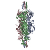

 Sample preparation
 Electron microscopy imaging
 FIELD EMISSION GUN / Accelerating voltage: 300 kV / Illumination mode: FLOOD BEAM
 Processing