[English] 日本語
Yorodumi
- PDB-7vy7: Snapshots of Human PSMD10(Gankyrin) unfolding by urea: 3 hours in... -

+
Open data


ID or keywords:

Loading...

-
Basic information

Entry
Database: PDB / ID: 7vy7
TitleSnapshots of Human PSMD10(Gankyrin) unfolding by urea: 3 hours incubation
Components26S proteasome non-ATPase regulatory subunit 10
KeywordsONCOPROTEIN / Proteasomal Chaperone / Urea denaturation
Function / homology
Function and homology information


proteasome regulatory particle assembly / positive regulation of cyclin-dependent protein serine/threonine kinase activity / Proteasome assembly / transcription factor binding / negative regulation of release of cytochrome c from mitochondria / negative regulation of NF-kappaB transcription factor activity / negative regulation of DNA damage response, signal transduction by p53 class mediator / negative regulation of MAPK cascade / proteasome complex / positive regulation of protein ubiquitination ...proteasome regulatory particle assembly / positive regulation of cyclin-dependent protein serine/threonine kinase activity / Proteasome assembly / transcription factor binding / negative regulation of release of cytochrome c from mitochondria / negative regulation of NF-kappaB transcription factor activity / negative regulation of DNA damage response, signal transduction by p53 class mediator / negative regulation of MAPK cascade / proteasome complex / positive regulation of protein ubiquitination / positive regulation of proteasomal ubiquitin-dependent protein catabolic process / microtubule cytoskeleton / positive regulation of cell growth / RNA polymerase II-specific DNA-binding transcription factor binding / cytoskeleton / cilium / apoptotic process / regulation of transcription by RNA polymerase II / negative regulation of apoptotic process / negative regulation of transcription by RNA polymerase II / nucleus / cytosol / cytoplasm
Similarity search - Function
: / Ankyrin repeat / Ankyrin repeats (3 copies) / Ankyrin repeat profile. / Ankyrin repeat region circular profile. / ankyrin repeats / Ankyrin repeat / Ankyrin repeat-containing domain superfamily
Similarity search - Domain/homology
UREA / 26S proteasome non-ATPase regulatory subunit 10
Similarity search - Component
Biological speciesHomo sapiens (human)
MethodX-RAY DIFFRACTION / MOLECULAR REPLACEMENT / molecular replacement / Resolution: 2.23 Å
AuthorsMukund Sudharsan, M.G. / Dalvi, S. / Venkatraman, P.
Funding support India, 1items
OrganizationGrant numberCountry
Other government5/13/62/2020-NCD-III India
CitationJournal: Protein Sci. / Year: 2022
Title: Snapshots of urea-induced early structural changes and unfolding of an ankyrin repeat protein at atomic resolution.
Authors: Medur Gurushankar, M.S. / Dalvi, S. / Venkatraman, P.
History
DepositionNov 13, 2021Deposition site: PDBJ / Processing site: PDBJ
Revision 1.0Nov 16, 2022Provider: repository / Type: Initial release
Revision 1.1Dec 28, 2022Group: Database references / Category: citation / citation_author
Item: _citation.country / _citation.journal_abbrev ..._citation.country / _citation.journal_abbrev / _citation.journal_id_ASTM / _citation.journal_id_CSD / _citation.journal_id_ISSN / _citation.journal_volume / _citation.page_first / _citation.page_last / _citation.pdbx_database_id_DOI / _citation.pdbx_database_id_PubMed / _citation.title / _citation.year / _citation_author.name
Revision 1.2Nov 29, 2023Group: Data collection / Refinement description
Category: chem_comp_atom / chem_comp_bond / pdbx_initial_refinement_model

-
Structure visualization

Structure viewerMolecule:
MolmilJmol/JSmol

Downloads & links

-
Assembly

Deposited unit
A: 26S proteasome non-ATPase regulatory subunit 10
hetero molecules


Theoretical massNumber of molelcules
Total (without water)24,6404
Polymers24,4601
Non-polymers1803
Water75742
1


  • Idetical with deposited unit
  • defined by author&software
TypeNameSymmetry operationNumber
identity operation1_555x,y,z1
Buried area190 Å2
ΔGint1 kcal/mol
Surface area10520 Å2
MethodPISA
Unit cell
Length a, b, c (Å)60.140, 60.140, 122.430
Angle α, β, γ (deg.)90.000, 90.000, 120.000
Int Tables number154
Space group name H-MP3221

-
Components

#1: Protein 26S proteasome non-ATPase regulatory subunit 10 / 26S proteasome regulatory subunit p28 / Gankyrin / p28(GANK)


Mass: 24459.855 Da / Num. of mol.: 1
Source method: isolated from a genetically manipulated source
Details: Gankyrin pBluescript II SK (1) construct (kind gift from Dr. Jun Fujita, Kyoto University)
Source: (gene. exp.) Homo sapiens (human) / Gene: PSMD10 / Plasmid: pRSETA / Production host: Escherichia coli (E. coli) / Strain (production host): Rosetta 2DE3 / References: UniProt: O75832
#2: Chemical ChemComp-URE / UREA


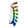
Mass: 60.055 Da / Num. of mol.: 3 / Source method: obtained synthetically / Formula: CH4N2O / Feature type: SUBJECT OF INVESTIGATION
#3: Water ChemComp-HOH / water


Mass: 18.015 Da / Num. of mol.: 42 / Source method: isolated from a natural source / Formula: H2O
Has ligand of interestY

-
Experimental details

-
Experiment

ExperimentMethod: X-RAY DIFFRACTION / Number of used crystals: 1

-
Sample preparation

CrystalDensity Matthews: 2.61 Å3/Da / Density % sol: 52.93 %
Crystal growTemperature: 295 K / Method: vapor diffusion, sitting drop / pH: 7 / Details: 60% Tacsimate pH 7

-
Data collection

DiffractionMean temperature: 100 K / Serial crystal experiment: N
Diffraction sourceSource: ROTATING ANODE / Type: BRUKER AXS MICROSTAR / Wavelength: 1.5418 Å
DetectorType: MAR scanner 345 mm plate / Detector: IMAGE PLATE / Date: Mar 9, 2021
RadiationMonochromator: M / Protocol: SINGLE WAVELENGTH / Monochromatic (M) / Laue (L): M / Scattering type: x-ray
Radiation wavelengthWavelength: 1.5418 Å / Relative weight: 1
ReflectionResolution: 2.228→52.083 Å / Num. all: 13085 / Num. obs: 13085 / % possible obs: 97.6 % / Redundancy: 8.3 % / Rpim(I) all: 0.096 / Rrim(I) all: 0.281 / Rsym value: 0.264 / Net I/av σ(I): 2.8 / Net I/σ(I): 7.4 / Num. measured all: 109128
Reflection shell

Diffraction-ID: 1

Resolution (Å)Redundancy (%)Rmerge(I) obsMean I/σ(I) obsNum. unique obsRpim(I) allRrim(I) allRsym value% possible all
2.228-2.338.11.3840.616020.5081.4771.38483.7
2.33-2.478.41.1650.718170.4231.241.165100
2.47-2.658.51.0310.716970.3731.0971.031100
2.65-2.868.50.6881.115930.2480.7320.688100
2.86-3.138.50.4971.514800.180.5290.497100
3.13-3.58.50.2543.113570.0920.270.254100
3.5-4.048.50.1275.911900.0460.1360.127100
4.04-4.958.40.0878.110350.0320.0930.087100
4.95-78.20.1165.78130.0420.1240.116100
7-52.0837.40.03915.45010.0150.0420.03999.9

-
Phasing

PhasingMethod: molecular replacement

-
Processing

Software
NameVersionClassificationNB
MOSFLMdata reduction
SCALA3.3.22data scaling
PHASERphasing
REFMAC5.8.0267refinement
PDB_EXTRACT3.27data extraction
RefinementMethod to determine structure: MOLECULAR REPLACEMENT
Starting model: 1UOH
Resolution: 2.23→52.08 Å / Cor.coef. Fo:Fc: 0.929 / Cor.coef. Fo:Fc free: 0.876 / SU B: 13.126 / SU ML: 0.29 / SU R Cruickshank DPI: 0.3151 / Cross valid method: THROUGHOUT / σ(F): 0 / ESU R: 0.315 / ESU R Free: 0.272 / Stereochemistry target values: MAXIMUM LIKELIHOOD
Details: HYDROGENS HAVE BEEN ADDED IN THE RIDING POSITIONS U VALUES : REFINED INDIVIDUALLY
RfactorNum. reflection% reflectionSelection details
Rfree0.3131 1307 10 %RANDOM
Rwork0.2396 ---
obs0.2469 11742 99.71 %-
Solvent computationIon probe radii: 0.8 Å / Shrinkage radii: 0.8 Å / VDW probe radii: 1.2 Å / Solvent model: MASK
Displacement parametersBiso max: 102.86 Å2 / Biso mean: 38.409 Å2 / Biso min: 14.94 Å2
Baniso -1Baniso -2Baniso -3
1-0.71 Å20.36 Å2-0 Å2
2--0.71 Å20 Å2
3----2.31 Å2
Refinement stepCycle: final / Resolution: 2.23→52.08 Å
ProteinNucleic acidLigandSolventTotal
Num. atoms1693 0 12 42 1747
Biso mean--39.6 31.24 -
Num. residues----222
Refine LS restraints
Refine-IDTypeDev idealDev ideal targetNumber
X-RAY DIFFRACTIONr_bond_refined_d0.0070.0131631
X-RAY DIFFRACTIONr_bond_other_d0.0010.0161546
X-RAY DIFFRACTIONr_angle_refined_deg1.6161.6222211
X-RAY DIFFRACTIONr_angle_other_deg2.9951.5783526
X-RAY DIFFRACTIONr_dihedral_angle_1_deg6.8765218
X-RAY DIFFRACTIONr_dihedral_angle_2_deg33.2724.24266
X-RAY DIFFRACTIONr_dihedral_angle_3_deg17.49715253
X-RAY DIFFRACTIONr_dihedral_angle_4_deg17.533155
X-RAY DIFFRACTIONr_chiral_restr0.0650.2224
X-RAY DIFFRACTIONr_gen_planes_refined0.0060.021884
X-RAY DIFFRACTIONr_gen_planes_other0.0010.02330
LS refinement shellResolution: 2.23→2.286 Å / Rfactor Rfree error: 0
RfactorNum. reflection% reflection
Rfree0.454 90 -
Rwork0.432 809 -
obs--96.15 %

+
About Yorodumi

-
News

-
Feb 9, 2022. New format data for meta-information of EMDB entries

New format data for meta-information of EMDB entries

  • Version 3 of the EMDB header file is now the official format.
  • The previous official version 1.9 will be removed from the archive.

Related info.:EMDB header

External links:wwPDB to switch to version 3 of the EMDB data model

-
Aug 12, 2020. Covid-19 info

Covid-19 info

URL: https://pdbj.org/emnavi/covid19.php

New page: Covid-19 featured information page in EM Navigator.

Related info.:Covid-19 info / Mar 5, 2020. Novel coronavirus structure data

+
Mar 5, 2020. Novel coronavirus structure data

Novel coronavirus structure data

Related info.:Yorodumi Speices / Aug 12, 2020. Covid-19 info

External links:COVID-19 featured content - PDBj / Molecule of the Month (242):Coronavirus Proteases

+
Jan 31, 2019. EMDB accession codes are about to change! (news from PDBe EMDB page)

EMDB accession codes are about to change! (news from PDBe EMDB page)

  • The allocation of 4 digits for EMDB accession codes will soon come to an end. Whilst these codes will remain in use, new EMDB accession codes will include an additional digit and will expand incrementally as the available range of codes is exhausted. The current 4-digit format prefixed with “EMD-” (i.e. EMD-XXXX) will advance to a 5-digit format (i.e. EMD-XXXXX), and so on. It is currently estimated that the 4-digit codes will be depleted around Spring 2019, at which point the 5-digit format will come into force.
  • The EM Navigator/Yorodumi systems omit the EMD- prefix.

Related info.:Q: What is EMD? / ID/Accession-code notation in Yorodumi/EM Navigator

External links:EMDB Accession Codes are Changing Soon! / Contact to PDBj

+
Jul 12, 2017. Major update of PDB

Major update of PDB

  • wwPDB released updated PDB data conforming to the new PDBx/mmCIF dictionary.
  • This is a major update changing the version number from 4 to 5, and with Remediation, in which all the entries are updated.
  • In this update, many items about electron microscopy experimental information are reorganized (e.g. em_software).
  • Now, EM Navigator and Yorodumi are based on the updated data.

External links:wwPDB Remediation / Enriched Model Files Conforming to OneDep Data Standards Now Available in the PDB FTP Archive

-
Yorodumi

Thousand views of thousand structures

  • Yorodumi is a browser for structure data from EMDB, PDB, SASBDB, etc.
  • This page is also the successor to EM Navigator detail page, and also detail information page/front-end page for Omokage search.
  • The word "yorodu" (or yorozu) is an old Japanese word meaning "ten thousand". "mi" (miru) is to see.

Related info.:EMDB / PDB / SASBDB / Comparison of 3 databanks / Yorodumi Search / Aug 31, 2016. New EM Navigator & Yorodumi / Yorodumi Papers / Jmol/JSmol / Function and homology information / Changes in new EM Navigator and Yorodumi

Read more