 Movie
Movie Controller

[English] 日本語
 Yorodumi
- PDB-7vwe: Human peroxisome proliferator-activated receptor (PPAR) delta lig... -
+ Open data

- Basic information
| Entry | Database: PDB / ID: 7vwe | ||||||
|---|---|---|---|---|---|---|---|
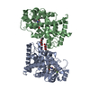
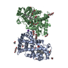
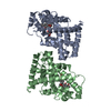
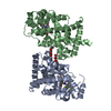
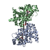
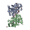
| Title | Human peroxisome proliferator-activated receptor (PPAR) delta ligand binding domain in complex with a synthetic partial agonist JK122 | ||||||
 Components Components | Peroxisome proliferator-activated receptor delta | ||||||
 Keywords Keywords | TRANSCRIPTION / NUCLEAR RECEPTOR / PROTEIN-LIGAND COMPLEX / PPAR | ||||||
| Function / homology |  Function and homology information Function and homology informationlinoleic acid binding / positive regulation of skeletal muscle tissue regeneration / positive regulation of epidermis development / axon ensheathment / regulation of skeletal muscle satellite cell proliferation / positive regulation of myoblast proliferation / D-glucose transmembrane transport / proteoglycan metabolic process / negative regulation of smooth muscle cell migration / fatty acid catabolic process ...linoleic acid binding / positive regulation of skeletal muscle tissue regeneration / positive regulation of epidermis development / axon ensheathment / regulation of skeletal muscle satellite cell proliferation / positive regulation of myoblast proliferation / D-glucose transmembrane transport / proteoglycan metabolic process / negative regulation of smooth muscle cell migration / fatty acid catabolic process / negative regulation of collagen biosynthetic process / negative regulation of myoblast differentiation / positive regulation of fatty acid oxidation / phospholipid biosynthetic process / Carnitine shuttle / response to vitamin A / Signaling by Retinoic Acid / positive regulation of fatty acid metabolic process / fatty acid beta-oxidation / negative regulation of cholesterol storage / nuclear steroid receptor activity / decidualization / response to glucose / positive regulation of fat cell differentiation / fatty acid transport / cholesterol metabolic process / NF-kappaB binding / energy homeostasis / embryo implantation / intracellular receptor signaling pathway / hormone-mediated signaling pathway / cellular response to nutrient levels / negative regulation of miRNA transcription / fatty acid metabolic process / positive regulation of insulin secretion involved in cellular response to glucose stimulus / response to activity / apoptotic signaling pathway / negative regulation of smooth muscle cell proliferation / lipid metabolic process / generation of precursor metabolites and energy / negative regulation of cell growth / Nuclear Receptor transcription pathway / negative regulation of inflammatory response / glucose metabolic process / vasodilation / cell population proliferation / DNA-binding transcription repressor activity, RNA polymerase II-specific / RNA polymerase II transcription regulator complex / nuclear receptor activity / transcription coactivator binding / sequence-specific double-stranded DNA binding / heart development / cellular response to lipopolysaccharide / DNA-binding transcription factor activity, RNA polymerase II-specific / cell differentiation / RNA polymerase II cis-regulatory region sequence-specific DNA binding / DNA-binding transcription factor activity / negative regulation of DNA-templated transcription / positive regulation of gene expression / apoptotic process / regulation of transcription by RNA polymerase II / lipid binding / negative regulation of apoptotic process / positive regulation of DNA-templated transcription / chromatin / negative regulation of transcription by RNA polymerase II / positive regulation of transcription by RNA polymerase II / DNA-templated transcription / DNA binding / nucleoplasm / zinc ion binding / nucleus Similarity search - Function | ||||||
| Biological species |  Homo sapiens (human) Homo sapiens (human) | ||||||
| Method |  X-RAY DIFFRACTION / SYNCHROTRON / MOLECULAR REPLACEMENT / Resolution: 3 Å X-RAY DIFFRACTION / SYNCHROTRON / MOLECULAR REPLACEMENT / Resolution: 3 Å | ||||||
 Authors Authors | Oyama, T. / Miyachi, H. | ||||||
| Funding support | 1items
| ||||||
 Citation Citation | Journal: Acta Crystallogr.,Sect.F / Year: 2022 Title: Crystal structures of the ligand-binding domain of human peroxisome proliferator-activated receptor delta in complexes with phenylpropanoic acid derivatives and a pyridine carboxylic acid derivative. Authors: Oyama, T. / Takiguchi, K. / Miyachi, H. | ||||||
| History |
|
- Structure visualization
| Structure viewer | Molecule: MolmilJmol/JSmol |
|---|
- Downloads & links
-Download
| PDBx/mmCIF format | 7vwe.cif.gz | 143.7 KB | Display | PDBx/mmCIF format |
|---|---|---|---|---|
| PDB format | pdb7vwe.ent.gz | 90.9 KB | Display | PDB format |
| PDBx/mmJSON format | 7vwe.json.gz | Tree view | PDBx/mmJSON format | |
| Others |  Other downloads Other downloads |
-Validation report
| Arichive directory | https://data.pdbj.org/pub/pdb/validation_reports/vw/7vweftp://data.pdbj.org/pub/pdb/validation_reports/vw/7vwe | HTTPS FTP |
|---|
-Related structure data
| Related structure data | 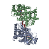 7vwfC 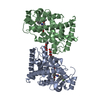 7vwgC  7vwhC  2znpS S: Starting model for refinement C: citing same article ( |
|---|---|
| Similar structure data |


-Links
 PDBj
PDBj
- Assembly
| Deposited unit | 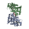
| ||||||||||||
|---|---|---|---|---|---|---|---|---|---|---|---|---|---|
| 1 |
| ||||||||||||
| Unit cell |
|
-Components
| #1: Protein | Mass: 31527.648 Da / Num. of mol.: 2 Source method: isolated from a genetically manipulated source Source: (gene. exp.) Homo sapiens (human) / Gene: PPARD, NR1C2, PPARB / Production host:  #2: Sugar |   Type: D-saccharide / Mass: 278.342 Da / Num. of mol.: 2 / Source method: obtained synthetically / Formula: C13H26O6 / Feature type: SUBJECT OF INVESTIGATION Type: D-saccharide / Mass: 278.342 Da / Num. of mol.: 2 / Source method: obtained synthetically / Formula: C13H26O6 / Feature type: SUBJECT OF INVESTIGATION#3: Chemical |   Mass: 518.500 Da / Num. of mol.: 2 / Source method: obtained synthetically / Formula: C27H26F4N2O4 / Feature type: SUBJECT OF INVESTIGATION Mass: 518.500 Da / Num. of mol.: 2 / Source method: obtained synthetically / Formula: C27H26F4N2O4 / Feature type: SUBJECT OF INVESTIGATION#4: Water | ChemComp-HOH / |  Mass: 18.015 Da / Num. of mol.: 9 / Source method: isolated from a natural source / Formula: H2O Mass: 18.015 Da / Num. of mol.: 9 / Source method: isolated from a natural source / Formula: H2OHas ligand of interest | Y | |
|---|
-Experimental details
-Experiment
| Experiment | Method: X-RAY DIFFRACTION / Number of used crystals: 1 |
|---|
- Sample preparation
| Crystal | Density Matthews: 2.72 Å3/Da / Density % sol: 54.85 % Description: THE ENTRY CONTAINS FRIEDEL PAIRS IN I/F_PLUS/MINUS COLUMNS. |
|---|---|
| Crystal grow | Temperature: 293 K / Method: vapor diffusion, hanging drop / pH: 9.5 Details: 22% Polyethyleneglycol, 200 mM potassium thiocyanate, 0.5% N-HEPTYL-BETA-D-GLUCOPYRANOSIDE |
-Data collection
| Diffraction | Mean temperature: 100 K / Serial crystal experiment: N |
|---|---|
| Diffraction source | Source: SYNCHROTRON / Site: Photon Factory  / Beamline: AR-NE3A / Wavelength: 1 Å / Beamline: AR-NE3A / Wavelength: 1 Å |
| Detector | Type: ADSC QUANTUM 270 / Detector: CCD / Date: Feb 11, 2013 |
| Radiation | Monochromator: SI(111) / Protocol: SINGLE WAVELENGTH / Monochromatic (M) / Laue (L): M / Scattering type: x-ray |
| Radiation wavelength | Wavelength: 1 Å / Relative weight: 1 |
| Reflection | Resolution: 3→50 Å / Num. obs: 26118 / % possible obs: 99.9 % / Redundancy: 3.7 % / Biso Wilson estimate: 57.18 Å2 / CC1/2: 0.996 / Rmerge(I) obs: 0.1 / Rpim(I) all: 0.06 / Rrim(I) all: 0.117 / Net I/σ(I): 7.9 |
| Reflection shell | Resolution: 3→3.18 Å / Redundancy: 3.7 % / Rmerge(I) obs: 0.48 / Num. unique obs: 2163 / CC1/2: 0.932 / Rpim(I) all: 0.287 / Rrim(I) all: 0.561 / % possible all: 99.9 |
- Processing
| Software |
| ||||||||||||||||||||||||||||||||||||||||||||||||||||||||||||||||||||||
|---|---|---|---|---|---|---|---|---|---|---|---|---|---|---|---|---|---|---|---|---|---|---|---|---|---|---|---|---|---|---|---|---|---|---|---|---|---|---|---|---|---|---|---|---|---|---|---|---|---|---|---|---|---|---|---|---|---|---|---|---|---|---|---|---|---|---|---|---|---|---|---|
| Refinement | Method to determine structure: MOLECULAR REPLACEMENT Starting model: 2znp Resolution: 3→47.57 Å / SU ML: 0.4532 / Cross valid method: FREE R-VALUE / σ(F): 0.2 / Phase error: 24.7644 Stereochemistry target values: GeoStd + Monomer Library + CDL v1.2
| ||||||||||||||||||||||||||||||||||||||||||||||||||||||||||||||||||||||
| Solvent computation | Shrinkage radii: 0.9 Å / VDW probe radii: 1.11 Å / Solvent model: FLAT BULK SOLVENT MODEL | ||||||||||||||||||||||||||||||||||||||||||||||||||||||||||||||||||||||
| Displacement parameters | Biso mean: 48.67 Å2 | ||||||||||||||||||||||||||||||||||||||||||||||||||||||||||||||||||||||
| Refinement step | Cycle: LAST / Resolution: 3→47.57 Å
| ||||||||||||||||||||||||||||||||||||||||||||||||||||||||||||||||||||||
| Refine LS restraints |
| ||||||||||||||||||||||||||||||||||||||||||||||||||||||||||||||||||||||
| LS refinement shell |
|
