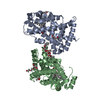
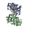
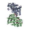
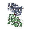
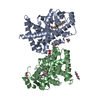
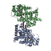
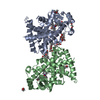
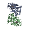
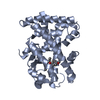
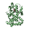
[English] 日本語
Yorodumi
- PDB-3d5f: Crystal Structure of PPAR-delta complex -

+
Open data


ID or keywords:

Loading...

-
Basic information

Entry
Database: PDB / ID: 3d5f
TitleCrystal Structure of PPAR-delta complex
ComponentsPeroxisome proliferator-activated receptor delta
KeywordsTRANSCRIPTION / Nuclear Receptor / Activator / Alternative splicing / DNA-binding / Metal-binding / Nucleus / Transcription regulation / Zinc / Zinc-finger
Function / homology
Function and homology information


fat cell proliferation / positive regulation of fat cell proliferation / keratinocyte migration / linoleic acid binding / positive regulation of skeletal muscle tissue regeneration / positive regulation of epidermis development / axon ensheathment / regulation of skeletal muscle satellite cell proliferation / D-glucose transmembrane transport / negative regulation of smooth muscle cell migration ...fat cell proliferation / positive regulation of fat cell proliferation / keratinocyte migration / linoleic acid binding / positive regulation of skeletal muscle tissue regeneration / positive regulation of epidermis development / axon ensheathment / regulation of skeletal muscle satellite cell proliferation / D-glucose transmembrane transport / negative regulation of smooth muscle cell migration / positive regulation of myoblast proliferation / proteoglycan metabolic process / fatty acid catabolic process / negative regulation of collagen biosynthetic process / positive regulation of fatty acid oxidation / phospholipid biosynthetic process / Carnitine shuttle / negative regulation of myoblast differentiation / response to vitamin A / Signaling by Retinoic Acid / positive regulation of fatty acid metabolic process / nuclear steroid receptor activity / fatty acid beta-oxidation / negative regulation of cholesterol storage / cell-substrate adhesion / positive regulation of insulin secretion involved in cellular response to glucose stimulus / decidualization / keratinocyte proliferation / positive regulation of fat cell differentiation / adipose tissue development / NF-kappaB binding / cellular response to nutrient levels / fatty acid transport / response to glucose / energy homeostasis / intracellular receptor signaling pathway / hormone-mediated signaling pathway / embryo implantation / cholesterol metabolic process / negative regulation of miRNA transcription / response to activity / fatty acid metabolic process / generation of precursor metabolites and energy / phosphatidylinositol 3-kinase/protein kinase B signal transduction / apoptotic signaling pathway / negative regulation of smooth muscle cell proliferation / wound healing / transcription coactivator binding / negative regulation of cell growth / Nuclear Receptor transcription pathway / lipid metabolic process / DNA-binding transcription repressor activity, RNA polymerase II-specific / negative regulation of inflammatory response / nuclear receptor activity / glucose metabolic process / negative regulation of epithelial cell proliferation / sequence-specific double-stranded DNA binding / vasodilation / heart development / cellular response to lipopolysaccharide / cellular response to hypoxia / cell differentiation / DNA-binding transcription factor activity, RNA polymerase II-specific / cell population proliferation / positive regulation of phosphatidylinositol 3-kinase/protein kinase B signal transduction / RNA polymerase II cis-regulatory region sequence-specific DNA binding / DNA-binding transcription factor activity / negative regulation of DNA-templated transcription / apoptotic process / lipid binding / positive regulation of gene expression / regulation of transcription by RNA polymerase II / negative regulation of apoptotic process / chromatin / positive regulation of DNA-templated transcription / negative regulation of transcription by RNA polymerase II / positive regulation of transcription by RNA polymerase II / DNA binding / zinc ion binding / nucleoplasm / nucleus
Similarity search - Function
Peroxisome proliferator-activated receptor, beta / Peroxisome proliferator-activated receptor / : / Retinoid X Receptor / Retinoid X Receptor / Nuclear hormone receptor / Nuclear hormones receptors DNA-binding region signature. / Zinc finger, nuclear hormone receptor-type / Double treble clef zinc finger, C4 type / Nuclear hormone receptors DNA-binding domain profile. ...Peroxisome proliferator-activated receptor, beta / Peroxisome proliferator-activated receptor / : / Retinoid X Receptor / Retinoid X Receptor / Nuclear hormone receptor / Nuclear hormones receptors DNA-binding region signature. / Zinc finger, nuclear hormone receptor-type / Double treble clef zinc finger, C4 type / Nuclear hormone receptors DNA-binding domain profile. / c4 zinc finger in nuclear hormone receptors / Nuclear hormone receptor, ligand-binding domain / Nuclear hormone receptor-like domain superfamily / Ligand-binding domain of nuclear hormone receptor / Nuclear receptor (NR) ligand-binding (LBD) domain profile. / Ligand binding domain of hormone receptors / Zinc finger, NHR/GATA-type / Orthogonal Bundle / Mainly Alpha
Similarity search - Domain/homology
Chem-L41 / Peroxisome proliferator-activated receptor delta
Similarity search - Component
Biological speciesHomo sapiens (human)
MethodX-RAY DIFFRACTION / SYNCHROTRON / MOLECULAR REPLACEMENT / Resolution: 2.2 Å
AuthorsAmano, Y.
CitationJournal: to be published
Title: PPAR-Delta Activation Contributes to Neuroprotectio Against Thapsigargin-Induced SH-SY5Y Cell Death
Authors: Iwashita, A. / Mihara, K. / Amano, Y. / Orita, M. / Matsuoka, N.
History
DepositionMay 16, 2008Deposition site: RCSB / Processing site: PDBJ
Revision 1.0Jun 3, 2008Provider: repository / Type: Initial release
Revision 1.1Jul 13, 2011Group: Version format compliance
Revision 1.2Nov 10, 2021Group: Database references / Derived calculations / Category: database_2 / struct_ref_seq_dif / struct_site
Item: _database_2.pdbx_DOI / _database_2.pdbx_database_accession ..._database_2.pdbx_DOI / _database_2.pdbx_database_accession / _struct_ref_seq_dif.details / _struct_site.pdbx_auth_asym_id / _struct_site.pdbx_auth_comp_id / _struct_site.pdbx_auth_seq_id
Revision 1.3Nov 1, 2023Group: Data collection / Refinement description
Category: chem_comp_atom / chem_comp_bond / pdbx_initial_refinement_model

-
Structure visualization

Structure viewerMolecule:
MolmilJmol/JSmol

Downloads & links

-
Assembly

Deposited unit
A: Peroxisome proliferator-activated receptor delta
B: Peroxisome proliferator-activated receptor delta
hetero molecules


Theoretical massNumber of molelcules
Total (without water)61,9424
Polymers61,1372
Non-polymers8052
Water1,56787
1
A: Peroxisome proliferator-activated receptor delta
hetero molecules

B: Peroxisome proliferator-activated receptor delta
hetero molecules


Theoretical massNumber of molelcules
Total (without water)61,9424
Polymers61,1372
Non-polymers8052
Water362
TypeNameSymmetry operationNumber
identity operation1_555x,y,z1
crystal symmetry operation2_656-x+1,y+1/2,-z+11
Buried area1370 Å2
ΔGint-15 kcal/mol
Surface area23330 Å2
MethodPISA
2
A: Peroxisome proliferator-activated receptor delta
hetero molecules


Theoretical massNumber of molelcules
Total (without water)30,9712
Polymers30,5691
Non-polymers4021
Water181
TypeNameSymmetry operationNumber
identity operation1_555x,y,z1
3
B: Peroxisome proliferator-activated receptor delta
hetero molecules


Theoretical massNumber of molelcules
Total (without water)30,9712
Polymers30,5691
Non-polymers4021
Water181
TypeNameSymmetry operationNumber
identity operation1_555x,y,z1
Unit cell
Length a, b, c (Å)39.456, 92.829, 96.454
Angle α, β, γ (deg.)90.000, 98.020, 90.000
Int Tables number4
Space group name H-MP1211

-
Components

#1: Protein Peroxisome proliferator-activated receptor delta / PPAR-delta / PPAR-beta / Nuclear receptor subfamily 1 group C member 2 / Nuclear hormone receptor 1 ...PPAR-delta / PPAR-beta / Nuclear receptor subfamily 1 group C member 2 / Nuclear hormone receptor 1 / NUC1 / NUCI


Mass: 30568.590 Da / Num. of mol.: 2 / Fragment: ligand binding domain, UNP residues 175-441 / Mutation: Y430Q
Source method: isolated from a genetically manipulated source
Source: (gene. exp.) Homo sapiens (human) / Plasmid: pET28 / Production host: Escherichia coli (E. coli) / Strain (production host): BL21(DE3) / References: UniProt: Q03181
#2: Chemical ChemComp-L41 / {4-[3-(4-acetyl-3-hydroxy-2-propylphenoxy)propoxy]phenoxy}acetic acid


Mass: 402.438 Da / Num. of mol.: 2 / Source method: obtained synthetically / Formula: C22H26O7 / Comment: agonist*YM
#3: Water ChemComp-HOH / water


Mass: 18.015 Da / Num. of mol.: 87 / Source method: isolated from a natural source / Formula: H2O

-
Experimental details

-
Experiment

ExperimentMethod: X-RAY DIFFRACTION / Number of used crystals: 1

-
Sample preparation

CrystalDensity Matthews: 2.86 Å3/Da / Density % sol: 57.01 %
Crystal growTemperature: 298 K / Method: vapor diffusion / pH: 6.5
Details: PEG 8000, pH 6.5, vapor diffusion, temperature 298K

-
Data collection

DiffractionMean temperature: 100 K
Diffraction sourceSource: SYNCHROTRON / Site: SPring-8 / Beamline: BL32B2 / Wavelength: 1 Å
DetectorType: RIGAKU JUPITER 210 / Detector: CCD / Date: Dec 20, 2004
RadiationProtocol: SINGLE WAVELENGTH / Monochromatic (M) / Laue (L): M / Scattering type: x-ray
Radiation wavelengthWavelength: 1 Å / Relative weight: 1
ReflectionResolution: 2.2→42.47 Å / Num. obs: 33944 / % possible obs: 97.1 % / Observed criterion σ(I): 3 / Redundancy: 3.84 % / Rmerge(I) obs: 0.129 / Rsym value: 0.129 / Net I/σ(I): 4.4
Reflection shellResolution: 2.2→2.28 Å / Redundancy: 3.81 % / Rmerge(I) obs: 0.416 / Mean I/σ(I) obs: 1.3 / Rsym value: 0.416 / % possible all: 99.2

-
Processing

Software
NameVersionClassificationNB
CNSrefinement
PDB_EXTRACT3.005data extraction
CrystalCleardata reduction
CrystalCleardata scaling
RefinementMethod to determine structure: MOLECULAR REPLACEMENT
Starting model: PDB ENTRY 1GWX
Resolution: 2.2→42.47 Å
RfactorNum. reflection% reflection
Rfree0.281 --
Rwork0.278 --
obs0.278 33944 96.9 %
Displacement parametersBiso mean: 38.641 Å2
Refinement stepCycle: LAST / Resolution: 2.2→42.47 Å
ProteinNucleic acidLigandSolventTotal
Num. atoms4100 0 58 87 4245

+
About Yorodumi

-
News

-
Feb 9, 2022. New format data for meta-information of EMDB entries

New format data for meta-information of EMDB entries

  • Version 3 of the EMDB header file is now the official format.
  • The previous official version 1.9 will be removed from the archive.

Related info.:EMDB header

External links:wwPDB to switch to version 3 of the EMDB data model

-
Aug 12, 2020. Covid-19 info

Covid-19 info

URL: https://pdbj.org/emnavi/covid19.php

New page: Covid-19 featured information page in EM Navigator.

Related info.:Covid-19 info / Mar 5, 2020. Novel coronavirus structure data

+
Mar 5, 2020. Novel coronavirus structure data

Novel coronavirus structure data

Related info.:Yorodumi Speices / Aug 12, 2020. Covid-19 info

External links:COVID-19 featured content - PDBj / Molecule of the Month (242):Coronavirus Proteases

+
Jan 31, 2019. EMDB accession codes are about to change! (news from PDBe EMDB page)

EMDB accession codes are about to change! (news from PDBe EMDB page)

  • The allocation of 4 digits for EMDB accession codes will soon come to an end. Whilst these codes will remain in use, new EMDB accession codes will include an additional digit and will expand incrementally as the available range of codes is exhausted. The current 4-digit format prefixed with “EMD-” (i.e. EMD-XXXX) will advance to a 5-digit format (i.e. EMD-XXXXX), and so on. It is currently estimated that the 4-digit codes will be depleted around Spring 2019, at which point the 5-digit format will come into force.
  • The EM Navigator/Yorodumi systems omit the EMD- prefix.

Related info.:Q: What is EMD? / ID/Accession-code notation in Yorodumi/EM Navigator

External links:EMDB Accession Codes are Changing Soon! / Contact to PDBj

+
Jul 12, 2017. Major update of PDB

Major update of PDB

  • wwPDB released updated PDB data conforming to the new PDBx/mmCIF dictionary.
  • This is a major update changing the version number from 4 to 5, and with Remediation, in which all the entries are updated.
  • In this update, many items about electron microscopy experimental information are reorganized (e.g. em_software).
  • Now, EM Navigator and Yorodumi are based on the updated data.

External links:wwPDB Remediation / Enriched Model Files Conforming to OneDep Data Standards Now Available in the PDB FTP Archive

-
Yorodumi

Thousand views of thousand structures

  • Yorodumi is a browser for structure data from EMDB, PDB, SASBDB, etc.
  • This page is also the successor to EM Navigator detail page, and also detail information page/front-end page for Omokage search.
  • The word "yorodu" (or yorozu) is an old Japanese word meaning "ten thousand". "mi" (miru) is to see.

Related info.:EMDB / PDB / SASBDB / Comparison of 3 databanks / Yorodumi Search / Aug 31, 2016. New EM Navigator & Yorodumi / Yorodumi Papers / Jmol/JSmol / Function and homology information / Changes in new EM Navigator and Yorodumi

Read more