 Movie
Movie Controller

[English] 日本語
 Yorodumi
- PDB-7vwb: Phloem lectin (PP2) structure -complex with N-Acetyllactosamine (... -
+ Open data

- Basic information
| Entry | Database: PDB / ID: 7vwb | ||||||
|---|---|---|---|---|---|---|---|
| Title | Phloem lectin (PP2) structure -complex with N-Acetyllactosamine (LacNAc) | ||||||
 Components Components | phloem lectin | ||||||
 Keywords Keywords | SUGAR BINDING PROTEIN / Phloem lectin / chitin-binding lectin | ||||||
| Function / homology | : / Phloem protein 2-like / Phloem protein 2 / carbohydrate binding / N-acetyl-alpha-lactosamine / 17 kDa phloem lectin Function and homology information Function and homology information | ||||||
| Biological species |  Cucumis sativus (cucumber) Cucumis sativus (cucumber) | ||||||
| Method |  X-RAY DIFFRACTION / MOLECULAR REPLACEMENT / Resolution: 1.9 Å X-RAY DIFFRACTION / MOLECULAR REPLACEMENT / Resolution: 1.9 Å | ||||||
 Authors Authors | Sivaji, N. / Bobbili, K.B. / Suguna, K. / Surolia, A. | ||||||
| Funding support |  India, 1items India, 1items
| ||||||
 Citation Citation | Journal: Structure / Year: 2023 Title: Structure and interactions of the phloem lectin (phloem protein 2) Cus17 from Cucumis sativus. Authors: Sivaji, N. / Bobbili, K.B. / Suguna, K. / Surolia, A. | ||||||
| History |
|
- Structure visualization
| Structure viewer | Molecule: MolmilJmol/JSmol |
|---|
- Downloads & links
-Download
| PDBx/mmCIF format | 7vwb.cif.gz | 82.5 KB | Display | PDBx/mmCIF format |
|---|---|---|---|---|
| PDB format | pdb7vwb.ent.gz | 58.6 KB | Display | PDB format |
| PDBx/mmJSON format | 7vwb.json.gz | Tree view | PDBx/mmJSON format | |
| Others |  Other downloads Other downloads |
-Validation report
| Arichive directory | https://data.pdbj.org/pub/pdb/validation_reports/vw/7vwbftp://data.pdbj.org/pub/pdb/validation_reports/vw/7vwb | HTTPS FTP |
|---|
-Related structure data
| Related structure data |  7vs6SC  7vubC  7w4bC S: Starting model for refinement C: citing same article ( |
|---|---|
| Similar structure data |
-Links
 PDBj
PDBj- Assembly
| Deposited unit | 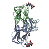
| ||||||||
|---|---|---|---|---|---|---|---|---|---|
| 1 |
| ||||||||
| Unit cell |
|
-Components
| #1: Protein | Mass: 17651.926 Da / Num. of mol.: 2 Source method: isolated from a genetically manipulated source Source: (gene. exp.) Cucumis sativus (cucumber) / Gene: Lec17-7 / Production host:  #2: Polysaccharide | 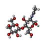  Source method: isolated from a genetically manipulated source Details: oligosaccharide / References: N-acetyl-alpha-lactosamine #3: Chemical | ChemComp-EDO / |   Mass: 62.068 Da / Num. of mol.: 1 / Source method: obtained synthetically / Formula: C2H6O2 Mass: 62.068 Da / Num. of mol.: 1 / Source method: obtained synthetically / Formula: C2H6O2#4: Water | ChemComp-HOH / |  Mass: 18.015 Da / Num. of mol.: 240 / Source method: isolated from a natural source / Formula: H2O Mass: 18.015 Da / Num. of mol.: 240 / Source method: isolated from a natural source / Formula: H2OHas ligand of interest | Y | Has protein modification | Y | |
|---|
-Experimental details
-Experiment
| Experiment | Method: X-RAY DIFFRACTION / Number of used crystals: 1 |
|---|
- Sample preparation
| Crystal | Density Matthews: 2.67 Å3/Da / Density % sol: 53.95 % |
|---|---|
| Crystal grow | Temperature: 295 K / Method: microbatch Details: 0.1 M MES monohydrate pH 6.0, 22% v/v Polyethylene glycol 400 |
-Data collection
| Diffraction | Mean temperature: 100 K / Serial crystal experiment: N |
|---|---|
| Diffraction source | Source: ROTATING ANODE / Type: RIGAKU ULTRAX 18 / Wavelength: 1.5418 Å |
| Detector | Type: RIGAKU RAXIS IV++ / Detector: IMAGE PLATE / Date: Feb 12, 2020 |
| Radiation | Protocol: SINGLE WAVELENGTH / Monochromatic (M) / Laue (L): M / Scattering type: x-ray |
| Radiation wavelength | Wavelength: 1.5418 Å / Relative weight: 1 |
| Reflection | Resolution: 1.9→30.19 Å / Num. obs: 28551 / % possible obs: 98 % / Redundancy: 4 % / CC1/2: 0.97 / Rmerge(I) obs: 0.13 / Net I/σ(I): 7 |
| Reflection shell | Resolution: 1.9→2 Å / Rmerge(I) obs: 0.18 / Num. unique obs: 4055 / CC1/2: 0.88 |
- Processing
| Software |
| |||||||||||||||||||||||||||||||||||||||||||||||||||||||||||||||||||||||||||||||||||||||||||||||||||||||||||||||||||||||||||||||||||||||||||||||||||||||||||||||||||||
|---|---|---|---|---|---|---|---|---|---|---|---|---|---|---|---|---|---|---|---|---|---|---|---|---|---|---|---|---|---|---|---|---|---|---|---|---|---|---|---|---|---|---|---|---|---|---|---|---|---|---|---|---|---|---|---|---|---|---|---|---|---|---|---|---|---|---|---|---|---|---|---|---|---|---|---|---|---|---|---|---|---|---|---|---|---|---|---|---|---|---|---|---|---|---|---|---|---|---|---|---|---|---|---|---|---|---|---|---|---|---|---|---|---|---|---|---|---|---|---|---|---|---|---|---|---|---|---|---|---|---|---|---|---|---|---|---|---|---|---|---|---|---|---|---|---|---|---|---|---|---|---|---|---|---|---|---|---|---|---|---|---|---|---|---|---|---|
| Refinement | Method to determine structure: MOLECULAR REPLACEMENT Starting model: 7VS6 Resolution: 1.9→24.896 Å / Cor.coef. Fo:Fc: 0.906 / Cor.coef. Fo:Fc free: 0.89 / Cross valid method: THROUGHOUT / ESU R: 0.174 / ESU R Free: 0.153 Details: Hydrogens have been added in their riding positions
| |||||||||||||||||||||||||||||||||||||||||||||||||||||||||||||||||||||||||||||||||||||||||||||||||||||||||||||||||||||||||||||||||||||||||||||||||||||||||||||||||||||
| Solvent computation | Ion probe radii: 0.8 Å / Shrinkage radii: 0.8 Å / VDW probe radii: 1.2 Å / Solvent model: MASK BULK SOLVENT | |||||||||||||||||||||||||||||||||||||||||||||||||||||||||||||||||||||||||||||||||||||||||||||||||||||||||||||||||||||||||||||||||||||||||||||||||||||||||||||||||||||
| Displacement parameters | Biso mean: 13.985 Å2
| |||||||||||||||||||||||||||||||||||||||||||||||||||||||||||||||||||||||||||||||||||||||||||||||||||||||||||||||||||||||||||||||||||||||||||||||||||||||||||||||||||||
| Refinement step | Cycle: LAST / Resolution: 1.9→24.896 Å
| |||||||||||||||||||||||||||||||||||||||||||||||||||||||||||||||||||||||||||||||||||||||||||||||||||||||||||||||||||||||||||||||||||||||||||||||||||||||||||||||||||||
| Refine LS restraints |
| |||||||||||||||||||||||||||||||||||||||||||||||||||||||||||||||||||||||||||||||||||||||||||||||||||||||||||||||||||||||||||||||||||||||||||||||||||||||||||||||||||||
| LS refinement shell |
|
