 Movie
Movie Controller


+ Open data

- Basic information
| Entry | Database: PDB / ID: 7vvy | ||||||
|---|---|---|---|---|---|---|---|
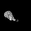
| Title | TRA module of NuA4 | ||||||
 Components Components |
| ||||||
 Keywords Keywords | DNA BINDING PROTEIN / NuA4 Tra1 | ||||||
| Function / homology |  Function and homology information Function and homology informationcellular bud neck contractile ring / mitotic actomyosin contractile ring contraction / RHOB GTPase cycle / RHOA GTPase cycle / piccolo histone acetyltransferase complex / vacuole inheritance / ascospore wall assembly / TTT Hsp90 cochaperone complex / actin cortical patch / SLIK (SAGA-like) complex ...cellular bud neck contractile ring / mitotic actomyosin contractile ring contraction / RHOB GTPase cycle / RHOA GTPase cycle / piccolo histone acetyltransferase complex / vacuole inheritance / ascospore wall assembly / TTT Hsp90 cochaperone complex / actin cortical patch / SLIK (SAGA-like) complex / kinetochore assembly / Swr1 complex / Ino80 complex / SAGA complex / SWI/SNF complex / DNA repair-dependent chromatin remodeling / actin filament bundle / establishment of cell polarity / NuA4 histone acetyltransferase complex / positive regulation of macroautophagy / protein secretion / chromosome organization / Ub-specific processing proteases / actin filament / structural constituent of cytoskeleton / Hydrolases; Acting on acid anhydrides; Acting on acid anhydrides to facilitate cellular and subcellular movement / endocytosis / transcription corepressor activity / nucleosome / actin cytoskeleton / chromatin organization / protein-containing complex assembly / histone binding / chromatin remodeling / DNA repair / DNA-templated transcription / chromatin binding / regulation of DNA-templated transcription / regulation of transcription by RNA polymerase II / chromatin / negative regulation of transcription by RNA polymerase II / positive regulation of transcription by RNA polymerase II / ATP hydrolysis activity / ATP binding / identical protein binding / nucleus Similarity search - Function | ||||||
| Biological species |  | ||||||
| Method | ELECTRON MICROSCOPY / single particle reconstruction / cryo EM / Resolution: 3.1 Å | ||||||
 Authors Authors | Chen, Z. / Qu, K. | ||||||
| Funding support | 1items
| ||||||
 Citation Citation | Journal: Nature / Year: 2022 Title: Structure of the NuA4 acetyltransferase complex bound to the nucleosome. Authors: Keke Qu / Kangjing Chen / Hao Wang / Xueming Li / Zhucheng Chen /  Abstract: Deoxyribonucleic acid in eukaryotes wraps around the histone octamer to form nucleosomes, the fundamental unit of chromatin. The N termini of histone H4 interact with nearby nucleosomes and play an ...Deoxyribonucleic acid in eukaryotes wraps around the histone octamer to form nucleosomes, the fundamental unit of chromatin. The N termini of histone H4 interact with nearby nucleosomes and play an important role in the formation of high-order chromatin structure and heterochromatin silencing. NuA4 in yeast and its homologue Tip60 complex in mammalian cells are the key enzymes that catalyse H4 acetylation, which in turn regulates chromatin packaging and function in transcription activation and DNA repair. Here we report the cryo-electron microscopy structure of NuA4 from Saccharomyces cerevisiae bound to the nucleosome. NuA4 comprises two major modules: the catalytic histone acetyltransferase (HAT) module and the transcription activator-binding (TRA) module. The nucleosome is mainly bound by the HAT module and is positioned close to a polybasic surface of the TRA module, which is important for the optimal activity of NuA4. The nucleosomal linker DNA carrying the upstream activation sequence is oriented towards the conserved, transcription activator-binding surface of the Tra1 subunit, which suggests a potential mechanism of NuA4 to act as a transcription co-activator. The HAT module recognizes the disk face of the nucleosome through the H2A-H2B acidic patch and nucleosomal DNA, projecting the catalytic pocket of Esa1 to the N-terminal tail of H4 and supporting its function in selective acetylation of H4. Together, our findings illustrate how NuA4 is assembled and provide mechanistic insights into nucleosome recognition and transcription co-activation by a HAT. | ||||||
| History |
|
- Structure visualization
| Structure viewer | Molecule: MolmilJmol/JSmol |
|---|
- Downloads & links
-Download
| PDBx/mmCIF format | 7vvy.cif.gz | 973.5 KB | Display | PDBx/mmCIF format |
|---|---|---|---|---|
| PDB format | pdb7vvy.ent.gz | 768.9 KB | Display | PDB format |
| PDBx/mmJSON format | 7vvy.json.gz | Tree view | PDBx/mmJSON format | |
| Others |  Other downloads Other downloads |
-Validation report
| Summary document | 7vvy_validation.pdf.gz | 1004.4 KB | Display | wwPDB validaton report |
|---|---|---|---|---|
| Full document | 7vvy_full_validation.pdf.gz | 1 MB | Display | |
| Data in XML | 7vvy_validation.xml.gz | 137.1 KB | Display | |
| Data in CIF | 7vvy_validation.cif.gz | 210.9 KB | Display | |
| Arichive directory | https://data.pdbj.org/pub/pdb/validation_reports/vv/7vvyftp://data.pdbj.org/pub/pdb/validation_reports/vv/7vvy | HTTPS FTP |
-Related structure data
| Related structure data | 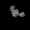 32149MC  7vvuC 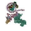 7vvzC M: map data used to model this data C: citing same article ( |
|---|---|
| Similar structure data |
-Links
 PDBj
PDBj


- Assembly
| Deposited unit | 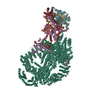
|
|---|---|
| 1 |
|
-Components
-Protein , 6 types, 6 molecules EFGKHL
| #1: Protein | Mass: 133949.953 Da / Num. of mol.: 1 Source method: isolated from a genetically manipulated source Source: (gene. exp.) |
|---|---|
| #2: Protein | Mass: 54894.684 Da / Num. of mol.: 1 Source method: isolated from a genetically manipulated source Source: (gene. exp.) |
| #3: Protein | Mass: 41735.547 Da / Num. of mol.: 1 Source method: isolated from a genetically manipulated source Source: (gene. exp.) |
| #4: Protein | Mass: 55297.684 Da / Num. of mol.: 1 Source method: isolated from a genetically manipulated source Source: (gene. exp.) |
| #5: Protein | Mass: 96889.867 Da / Num. of mol.: 1 Source method: isolated from a genetically manipulated source Source: (gene. exp.) |
| #6: Protein | Mass: 433677.281 Da / Num. of mol.: 1 Source method: isolated from a genetically manipulated source Source: (gene. exp.) Production host: |
-Non-polymers , 2 types, 4 molecules 

| #7: Chemical |  Mass: 24.305 Da / Num. of mol.: 2 / Source method: obtained synthetically / Formula: Mg / Feature type: SUBJECT OF INVESTIGATION Mass: 24.305 Da / Num. of mol.: 2 / Source method: obtained synthetically / Formula: Mg / Feature type: SUBJECT OF INVESTIGATION#8: Chemical |  Mass: 507.181 Da / Num. of mol.: 2 / Source method: obtained synthetically / Formula: C10H16N5O13P3 / Feature type: SUBJECT OF INVESTIGATION / Comment: ATP, energy-carrying molecule*YM Mass: 507.181 Da / Num. of mol.: 2 / Source method: obtained synthetically / Formula: C10H16N5O13P3 / Feature type: SUBJECT OF INVESTIGATION / Comment: ATP, energy-carrying molecule*YM |
|---|
-Details
| Has ligand of interest | Y |
|---|
-Experimental details
-Experiment
| Experiment | Method: ELECTRON MICROSCOPY |
|---|---|
| EM experiment | Aggregation state: PARTICLE / 3D reconstruction method: single particle reconstruction |
- Sample preparation
| Component | Name: TRA1 module of NuA4 / Type: COMPLEX / Entity ID: #1-#6 / Source: RECOMBINANT |
|---|---|
| Source (natural) | Organism: |
| Source (recombinant) | Organism: |
| Buffer solution | pH: 7.6 |
| Specimen | Embedding applied: NO / Shadowing applied: NO / Staining applied: NO / Vitrification applied: YES |
| Vitrification | Cryogen name: NITROGEN |
- Electron microscopy imaging
| Microscopy | Model: FEI TITAN |
|---|---|
| Electron gun | Electron source:  FIELD EMISSION GUN / Accelerating voltage: 300 kV / Illumination mode: FLOOD BEAM FIELD EMISSION GUN / Accelerating voltage: 300 kV / Illumination mode: FLOOD BEAM |
| Electron lens | Mode: BRIGHT FIELD / Nominal defocus max: 1800 nm / Nominal defocus min: 1300 nm |
| Image recording | Electron dose: 50 e/Å2 / Film or detector model: GATAN K3 (6k x 4k) |
- Processing
| CTF correction | Type: NONE |
|---|---|
| 3D reconstruction | Resolution: 3.1 Å / Resolution method: FSC 0.143 CUT-OFF / Num. of particles: 69770 / Symmetry type: POINT |
