 Movie
Movie Controller

[English] 日本語
 Yorodumi
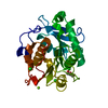
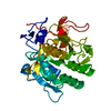
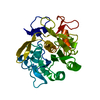
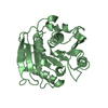
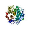
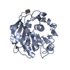
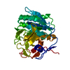
- PDB-7vvc: Crystal structure of inactive mutant of leaf-branch compost cutin... -
+ Open data

- Basic information
| Entry | Database: PDB / ID: 7vvc | ||||||
|---|---|---|---|---|---|---|---|
| Title | Crystal structure of inactive mutant of leaf-branch compost cutinase variant | ||||||
 Components Components | Leaf-branch compost cutinase | ||||||
 Keywords Keywords | HYDROLASE / plastic degradation / activity | ||||||
| Function / homology |  Function and homology information Function and homology informationacetylesterase activity / poly(ethylene terephthalate) hydrolase / cutinase / cutinase activity / extracellular region Similarity search - Function | ||||||
| Biological species |  Unknown prokaryotic organism (environmental samples) Unknown prokaryotic organism (environmental samples) | ||||||
| Method |  X-RAY DIFFRACTION / MOLECULAR REPLACEMENT / Resolution: 1.82 Å X-RAY DIFFRACTION / MOLECULAR REPLACEMENT / Resolution: 1.82 Å | ||||||
 Authors Authors | Niu, D. / Zeng, W. / Huang, J.W. / Chen, C.C. / Liu, W.D. / Guo, R.T. | ||||||
| Funding support | 1items
| ||||||
 Citation Citation | Journal: Acs Catalysis / Year: 2022 Title: Substrate-Binding Mode of a Thermophilic PET Hydrolase and Engineering the Enzyme to Enhance the Hydrolytic Efficacy. Authors: Zeng, W. / Li, X. / Yang, Y. / Min, J. / Huang, J.-W. / Liu, W. / Niu, D. / Yang, X. / Han, X. / Zhang, L. / Dai, L. / Chen, C.-C. / Guo, R.-T. | ||||||
| History |
|
- Structure visualization
| Structure viewer | Molecule: MolmilJmol/JSmol |
|---|
- Downloads & links
-Download
| PDBx/mmCIF format | 7vvc.cif.gz | 131 KB | Display | PDBx/mmCIF format |
|---|---|---|---|---|
| PDB format | pdb7vvc.ent.gz | 98.2 KB | Display | PDB format |
| PDBx/mmJSON format | 7vvc.json.gz | Tree view | PDBx/mmJSON format | |
| Others |  Other downloads Other downloads |
-Validation report
| Arichive directory | https://data.pdbj.org/pub/pdb/validation_reports/vv/7vvcftp://data.pdbj.org/pub/pdb/validation_reports/vv/7vvc | HTTPS FTP |
|---|
-Related structure data
| Related structure data | 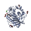 7vveC 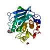 7w1nC 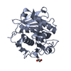 7w44C 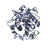 7w45C 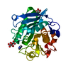 7ds7S S: Starting model for refinement C: citing same article ( |
|---|---|
| Similar structure data |


-Links
 PDBj
PDBj


- Assembly
| Deposited unit | 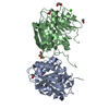
| ||||||||
|---|---|---|---|---|---|---|---|---|---|
| 1 | 
| ||||||||
| 2 | 
| ||||||||
| Unit cell |
|
-Components
-Protein , 1 types, 2 molecules AB
| #1: Protein | Mass: 28476.861 Da / Num. of mol.: 2 Source method: isolated from a genetically manipulated source Source: (gene. exp.) Unknown prokaryotic organism (environmental samples)Plasmid: pET32a / Production host: References: UniProt: G9BY57, cutinase, poly(ethylene terephthalate) hydrolase |
|---|
-Non-polymers , 5 types, 724 molecules 


| #2: Chemical |  Mass: 40.078 Da / Num. of mol.: 2 / Source method: obtained synthetically / Formula: Ca Mass: 40.078 Da / Num. of mol.: 2 / Source method: obtained synthetically / Formula: Ca#3: Chemical |  Mass: 60.052 Da / Num. of mol.: 2 / Source method: obtained synthetically / Formula: C2H4O2 Mass: 60.052 Da / Num. of mol.: 2 / Source method: obtained synthetically / Formula: C2H4O2#4: Chemical |  Mass: 92.094 Da / Num. of mol.: 2 / Source method: obtained synthetically / Formula: C3H8O3 Mass: 92.094 Da / Num. of mol.: 2 / Source method: obtained synthetically / Formula: C3H8O3#5: Chemical | ChemComp-ACT / |  Mass: 59.044 Da / Num. of mol.: 1 / Source method: obtained synthetically / Formula: C2H3O2 Mass: 59.044 Da / Num. of mol.: 1 / Source method: obtained synthetically / Formula: C2H3O2#6: Water | ChemComp-HOH / | Mass: 18.015 Da / Num. of mol.: 717 / Source method: isolated from a natural source / Formula: H2O |
|---|
-Details
| Has ligand of interest | N |
|---|---|
| Has protein modification | Y |
-Experimental details
-Experiment
| Experiment | Method: X-RAY DIFFRACTION / Number of used crystals: 1 |
|---|
- Sample preparation
| Crystal | Density Matthews: 2.27 Å3/Da / Density % sol: 45.82 % / Mosaicity: 0 ° |
|---|---|
| Crystal grow | Temperature: 298 K / Method: vapor diffusion, sitting drop / pH: 4.6 Details: Ammonium Acetate,Sodium Acetate trihydrate,Polyethylene Glycol 4000, Glycerol |
-Data collection
| Diffraction | Mean temperature: 100 K / Serial crystal experiment: N |
|---|---|
| Diffraction source | Source: LIQUID ANODE / Type: BRUKER METALJET / Wavelength: 1.34138 Å |
| Detector | Type: Bruker PHOTON III / Detector: PIXEL / Date: Jul 8, 2021 |
| Radiation | Protocol: SINGLE WAVELENGTH / Monochromatic (M) / Laue (L): M / Scattering type: x-ray |
| Radiation wavelength | Wavelength: 1.34138 Å / Relative weight: 1 |
| Reflection | Resolution: 1.82→25 Å / Num. obs: 45015 / % possible obs: 99.8 % / Redundancy: 4.8 % / CC1/2: 0.997 / Rmerge(I) obs: 0.076 / Rpim(I) all: 0.038 / Rrim(I) all: 0.085 / Net I/σ(I): 12.4 |
| Reflection shell | Resolution: 1.85→1.89 Å / Redundancy: 3.5 % / Rmerge(I) obs: 0.25 / Num. unique obs: 2722 / CC1/2: 0.952 / Rpim(I) all: 0.155 / Rrim(I) all: 0.295 / % possible all: 99.2 |
- Processing
| Software |
| ||||||||||||||||||||||||||||||||||||||||||||||||||||||||||||
|---|---|---|---|---|---|---|---|---|---|---|---|---|---|---|---|---|---|---|---|---|---|---|---|---|---|---|---|---|---|---|---|---|---|---|---|---|---|---|---|---|---|---|---|---|---|---|---|---|---|---|---|---|---|---|---|---|---|---|---|---|---|
| Refinement | Method to determine structure: MOLECULAR REPLACEMENT Starting model: 7DS7 Resolution: 1.82→25 Å / Cor.coef. Fo:Fc: 0.966 / Cor.coef. Fo:Fc free: 0.942 / SU B: 2.718 / SU ML: 0.082 / Cross valid method: THROUGHOUT / σ(F): 0 / ESU R: 0.122 / ESU R Free: 0.12 / Stereochemistry target values: MAXIMUM LIKELIHOOD Details: HYDROGENS HAVE BEEN ADDED IN THE RIDING POSITIONS U VALUES : REFINED INDIVIDUALLY
| ||||||||||||||||||||||||||||||||||||||||||||||||||||||||||||
| Solvent computation | Ion probe radii: 0.8 Å / Shrinkage radii: 0.8 Å / VDW probe radii: 1.2 Å / Solvent model: MASK | ||||||||||||||||||||||||||||||||||||||||||||||||||||||||||||
| Displacement parameters | Biso max: 64.52 Å2 / Biso mean: 14.49 Å2 / Biso min: 5.56 Å2
| ||||||||||||||||||||||||||||||||||||||||||||||||||||||||||||
| Refinement step | Cycle: final / Resolution: 1.82→25 Å
| ||||||||||||||||||||||||||||||||||||||||||||||||||||||||||||
| Refine LS restraints |
| ||||||||||||||||||||||||||||||||||||||||||||||||||||||||||||
| LS refinement shell | Resolution: 1.82→1.867 Å / Rfactor Rfree error: 0 / Total num. of bins used: 20
|
