 Movie
Movie Controller

[English] 日本語
 Yorodumi
- PDB-7vtd: Unliganded structure of Pseudouridine kinase (PUKI) from Escheric... -
+ Open data

- Basic information
| Entry | Database: PDB / ID: 7vtd | ||||||
|---|---|---|---|---|---|---|---|
| Title | Unliganded structure of Pseudouridine kinase (PUKI) from Escherichia coli strain B | ||||||
 Components Components | Pseudouridine kinase | ||||||
 Keywords Keywords | TRANSFERASE / pseudouridine kinase / pseudourdine / pfkb family | ||||||
| Function / homology |  Function and homology information Function and homology information | ||||||
| Biological species |  | ||||||
| Method |  X-RAY DIFFRACTION / SYNCHROTRON / MOLECULAR REPLACEMENT / Resolution: 2.15054869646 Å X-RAY DIFFRACTION / SYNCHROTRON / MOLECULAR REPLACEMENT / Resolution: 2.15054869646 Å | ||||||
 Authors Authors | Kim, S.H. / Rhee, S. | ||||||
| Funding support |  Korea, Republic Of, 1items Korea, Republic Of, 1items
| ||||||
 Citation Citation | Journal: J.Biol.Chem. / Year: 2022 Title: Substrate-binding loop interactions with pseudouridine trigger conformational changes that promote catalytic efficiency of pseudouridine kinase PUKI. Authors: Kim, S.H. / Kim, M. / Park, D. / Byun, S. / Rhee, S. | ||||||
| History |
|
- Structure visualization
| Structure viewer | Molecule: MolmilJmol/JSmol |
|---|
- Downloads & links
-Download
| PDBx/mmCIF format | 7vtd.cif.gz | 564 KB | Display | PDBx/mmCIF format |
|---|---|---|---|---|
| PDB format | pdb7vtd.ent.gz | 390.1 KB | Display | PDB format |
| PDBx/mmJSON format | 7vtd.json.gz | Tree view | PDBx/mmJSON format | |
| Others |  Other downloads Other downloads |
-Validation report
| Arichive directory | https://data.pdbj.org/pub/pdb/validation_reports/vt/7vtdftp://data.pdbj.org/pub/pdb/validation_reports/vt/7vtd | HTTPS FTP |
|---|
-Related structure data
| Related structure data |  7vteC  7vtfC  7vtgC  7vvaC  3kzhS S: Starting model for refinement C: citing same article ( |
|---|---|
| Similar structure data |
-Links
 PDBj
PDBj- Assembly
| Deposited unit | 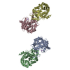
| ||||||||||||||||||||||||||||||
|---|---|---|---|---|---|---|---|---|---|---|---|---|---|---|---|---|---|---|---|---|---|---|---|---|---|---|---|---|---|---|---|
| 1 | 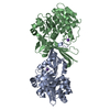
| ||||||||||||||||||||||||||||||
| 2 | 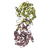
| ||||||||||||||||||||||||||||||
| Unit cell |
| ||||||||||||||||||||||||||||||
| Noncrystallographic symmetry (NCS) | NCS domain:
NCS domain segments: Component-ID: 1 / Ens-ID: 1 / Beg auth comp-ID: GLU / Beg label comp-ID: GLU / End auth comp-ID: ASN / End label comp-ID: ASN / Auth seq-ID: 3 - 308 / Label seq-ID: 3 - 308
|
-Components
| #1: Protein | Mass: 33781.094 Da / Num. of mol.: 4 Source method: isolated from a genetically manipulated source Source: (gene. exp.) Gene: psuK, psuK_1, psuK_2, A8C65_16945, BMT91_00230, BON75_04205, BvCmsHHP019_05067, BvCmsSINP011_02211, D9H68_16155, D9J11_19520, E2127_01880, E2128_04080, E2134_03530, FORC82_1677, G9P50_07470, ...Gene: psuK, psuK_1, psuK_2, A8C65_16945, BMT91_00230, BON75_04205, BvCmsHHP019_05067, BvCmsSINP011_02211, D9H68_16155, D9J11_19520, E2127_01880, E2128_04080, E2134_03530, FORC82_1677, G9P50_07470, GQE64_02140, GQM28_02110, HVV70_14180, HVX33_21995, HVZ33_08660, NCTC11022_02147, NCTC13216_04879, NCTC9706_04998, SAMEA3752557_00486, WP4S18E08_16210 Production host: #2: Chemical | ChemComp-K /   Mass: 39.098 Da / Num. of mol.: 4 / Source method: obtained synthetically / Formula: K Mass: 39.098 Da / Num. of mol.: 4 / Source method: obtained synthetically / Formula: K#3: Water | ChemComp-HOH / |  Mass: 18.015 Da / Num. of mol.: 204 / Source method: isolated from a natural source / Formula: H2O Mass: 18.015 Da / Num. of mol.: 204 / Source method: isolated from a natural source / Formula: H2OHas ligand of interest | N | |
|---|
-Experimental details
-Experiment
| Experiment | Method: X-RAY DIFFRACTION / Number of used crystals: 1 |
|---|
- Sample preparation
| Crystal | Density Matthews: 3.7 Å3/Da / Density % sol: 66.77 % |
|---|---|
| Crystal grow | Temperature: 295 K / Method: vapor diffusion, hanging drop / Details: 0.2M magnesium formate, 20% PEG 3350, 5% glycerol |
-Data collection
| Diffraction | Mean temperature: 100 K / Serial crystal experiment: N |
|---|---|
| Diffraction source | Source: SYNCHROTRON / Site: PAL/PLS / Beamline: 7A (6B, 6C1) / Wavelength: 0.97934 Å |
| Detector | Type: ADSC QUANTUM 270 / Detector: CCD / Date: Nov 11, 2011 |
| Radiation | Protocol: SINGLE WAVELENGTH / Monochromatic (M) / Laue (L): M / Scattering type: x-ray |
| Radiation wavelength | Wavelength: 0.97934 Å / Relative weight: 1 |
| Reflection | Resolution: 2.15→50 Å / Num. obs: 104841 / % possible obs: 99.7 % / Redundancy: 5.4 % / Biso Wilson estimate: 49.5724471261 Å2 / CC1/2: 0.99 / Net I/σ(I): 15 |
| Reflection shell | Resolution: 2.15→2.23 Å / Num. unique obs: 10500 / CC1/2: 0.5 |
- Processing
| Software |
| |||||||||||||||||||||||||||||||||||||||||||||||||||||||||||||||||||||||||||||||||||||||||||||||||||||||||
|---|---|---|---|---|---|---|---|---|---|---|---|---|---|---|---|---|---|---|---|---|---|---|---|---|---|---|---|---|---|---|---|---|---|---|---|---|---|---|---|---|---|---|---|---|---|---|---|---|---|---|---|---|---|---|---|---|---|---|---|---|---|---|---|---|---|---|---|---|---|---|---|---|---|---|---|---|---|---|---|---|---|---|---|---|---|---|---|---|---|---|---|---|---|---|---|---|---|---|---|---|---|---|---|---|---|---|
| Refinement | Method to determine structure: MOLECULAR REPLACEMENT Starting model: 3KZH Resolution: 2.15054869646→33.2888937491 Å / SU ML: 0.507065525363 / Cross valid method: FREE R-VALUE / σ(F): 1.95834498789 / Phase error: 41.2002494246
| |||||||||||||||||||||||||||||||||||||||||||||||||||||||||||||||||||||||||||||||||||||||||||||||||||||||||
| Solvent computation | Shrinkage radii: 0.9 Å / VDW probe radii: 1.11 Å / Solvent model: FLAT BULK SOLVENT MODEL | |||||||||||||||||||||||||||||||||||||||||||||||||||||||||||||||||||||||||||||||||||||||||||||||||||||||||
| Displacement parameters | Biso mean: 85.9681233502 Å2 | |||||||||||||||||||||||||||||||||||||||||||||||||||||||||||||||||||||||||||||||||||||||||||||||||||||||||
| Refinement step | Cycle: LAST / Resolution: 2.15054869646→33.2888937491 Å
| |||||||||||||||||||||||||||||||||||||||||||||||||||||||||||||||||||||||||||||||||||||||||||||||||||||||||
| Refine LS restraints |
| |||||||||||||||||||||||||||||||||||||||||||||||||||||||||||||||||||||||||||||||||||||||||||||||||||||||||
| LS refinement shell |
| |||||||||||||||||||||||||||||||||||||||||||||||||||||||||||||||||||||||||||||||||||||||||||||||||||||||||
| Refinement TLS params. | Method: refined / Origin x: 61.0291501445 Å / Origin y: -1.80495814318 Å / Origin z: 14.4077919991 Å
| |||||||||||||||||||||||||||||||||||||||||||||||||||||||||||||||||||||||||||||||||||||||||||||||||||||||||
| Refinement TLS group | Selection details: all |
