 Movie
Movie Controller


+ Open data

- Basic information
| Entry | Database: PDB / ID: 7vt8 | ||||||
|---|---|---|---|---|---|---|---|
| Title | Crystal structure of MtGlu5 from Meiothermus taiwanensis WR-220 | ||||||
 Components Components | Endoglucanase H | ||||||
 Keywords Keywords | HYDROLASE / cellulase / GH5 / Meiothermus taiwanensis WR-220 / endo-beta-1 / 4-glucanase | ||||||
| Function / homology |  Function and homology information Function and homology informationglucan catabolic process / cellulase / cellulase activity / beta-glucosidase activity / cell surface / extracellular region Similarity search - Function | ||||||
| Biological species |  Meiothermus taiwanensis WR-220 (bacteria) Meiothermus taiwanensis WR-220 (bacteria) | ||||||
| Method |  X-RAY DIFFRACTION / SYNCHROTRON / MOLECULAR REPLACEMENT / Resolution: 2.99 Å X-RAY DIFFRACTION / SYNCHROTRON / MOLECULAR REPLACEMENT / Resolution: 2.99 Å | ||||||
 Authors Authors | Ye, T.J. / Ko, P.T. / Huang, K.F. / Wu, S.H. | ||||||
| Funding support |  Taiwan, 1items Taiwan, 1items
| ||||||
 Citation Citation | Journal: Acta Crystallogr D Struct Biol / Year: 2022 Title: Synergic action of an inserted carbohydrate-binding module in a glycoside hydrolase family 5 endoglucanase. Authors: Ye, T.J. / Huang, K.F. / Ko, T.P. / Wu, S.H. | ||||||
| History |
|
- Structure visualization
| Structure viewer | Molecule: MolmilJmol/JSmol |
|---|
- Downloads & links
-Download
| PDBx/mmCIF format | 7vt8.cif.gz | 194.7 KB | Display | PDBx/mmCIF format |
|---|---|---|---|---|
| PDB format | pdb7vt8.ent.gz | 156.3 KB | Display | PDB format |
| PDBx/mmJSON format | 7vt8.json.gz | Tree view | PDBx/mmJSON format | |
| Others |  Other downloads Other downloads |
-Validation report
| Arichive directory | https://data.pdbj.org/pub/pdb/validation_reports/vt/7vt8ftp://data.pdbj.org/pub/pdb/validation_reports/vt/7vt8 | HTTPS FTP |
|---|
-Related structure data
| Related structure data | 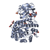 7vt4C 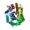 7vt5C 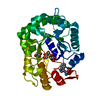 7vt6C 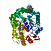 7vt7C  3mmuS S: Starting model for refinement C: citing same article ( |
|---|---|
| Similar structure data |
-Links
 PDBj
PDBj


- Assembly
| Deposited unit | 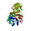
| ||||||||
|---|---|---|---|---|---|---|---|---|---|
| 1 |
| ||||||||
| Unit cell |
|
-Components
| #1: Protein | Mass: 52977.117 Da / Num. of mol.: 1 Source method: isolated from a genetically manipulated source Source: (gene. exp.) Meiothermus taiwanensis WR-220 (bacteria)Production host: | ||||
|---|---|---|---|---|---|
| #2: Sugar | ChemComp-BGC /   Type: D-saccharide, beta linking / Mass: 180.156 Da / Num. of mol.: 1 / Source method: obtained synthetically / Formula: C6H12O6 / Feature type: SUBJECT OF INVESTIGATION Type: D-saccharide, beta linking / Mass: 180.156 Da / Num. of mol.: 1 / Source method: obtained synthetically / Formula: C6H12O6 / Feature type: SUBJECT OF INVESTIGATION | ||||
| #3: Chemical | ChemComp-SO4 /   Mass: 96.063 Da / Num. of mol.: 8 / Source method: obtained synthetically / Formula: SO4 / Feature type: SUBJECT OF INVESTIGATION Mass: 96.063 Da / Num. of mol.: 8 / Source method: obtained synthetically / Formula: SO4 / Feature type: SUBJECT OF INVESTIGATION#4: Water | ChemComp-HOH / |  Mass: 18.015 Da / Num. of mol.: 36 / Source method: isolated from a natural source / Formula: H2O Mass: 18.015 Da / Num. of mol.: 36 / Source method: isolated from a natural source / Formula: H2OHas ligand of interest | Y | |
-Experimental details
-Experiment
| Experiment | Method: X-RAY DIFFRACTION / Number of used crystals: 1 |
|---|
- Sample preparation
| Crystal | Density Matthews: 4.9 Å3/Da / Density % sol: 74.88 % |
|---|---|
| Crystal grow | Temperature: 298 K / Method: microbatch Details: 0.16M citric acid, 1.1M ammonium sulfate, 10% PEG-400, pH 5.0 |
-Data collection
| Diffraction | Mean temperature: 100 K / Serial crystal experiment: N |
|---|---|
| Diffraction source | Source: SYNCHROTRON / Site: NSRRC / Beamline: TPS 05A / Wavelength: 1 Å |
| Detector | Type: RAYONIX MX300-HS / Detector: CCD / Date: Apr 11, 2021 |
| Radiation | Protocol: SINGLE WAVELENGTH / Monochromatic (M) / Laue (L): M / Scattering type: x-ray |
| Radiation wavelength | Wavelength: 1 Å / Relative weight: 1 |
| Reflection | Resolution: 2.99→50 Å / Num. obs: 21603 / % possible obs: 99.8 % / Redundancy: 7.8 % / Rmerge(I) obs: 0.071 / Rpim(I) all: 0.027 / Rrim(I) all: 0.076 / Χ2: 0.781 / Net I/σ(I): 26.55 |
| Reflection shell | Resolution: 2.99→3.04 Å / Num. unique obs: 1055 / CC1/2: 0.866 / Rpim(I) all: 0.439 / Χ2: 0.657 |
- Processing
| Software |
| |||||||||||||||||||||||||||||||||||||||||||||||||||||||||||||||||||||||||||||||||||||||||||||||||||||||||||||||||||||||||||||
|---|---|---|---|---|---|---|---|---|---|---|---|---|---|---|---|---|---|---|---|---|---|---|---|---|---|---|---|---|---|---|---|---|---|---|---|---|---|---|---|---|---|---|---|---|---|---|---|---|---|---|---|---|---|---|---|---|---|---|---|---|---|---|---|---|---|---|---|---|---|---|---|---|---|---|---|---|---|---|---|---|---|---|---|---|---|---|---|---|---|---|---|---|---|---|---|---|---|---|---|---|---|---|---|---|---|---|---|---|---|---|---|---|---|---|---|---|---|---|---|---|---|---|---|---|---|---|
| Refinement | Method to determine structure: MOLECULAR REPLACEMENT Starting model: 3mmu Resolution: 2.99→29.21 Å / SU ML: 0.43 / Cross valid method: THROUGHOUT / σ(F): 1.34 / Phase error: 31.52 / Stereochemistry target values: ML
| |||||||||||||||||||||||||||||||||||||||||||||||||||||||||||||||||||||||||||||||||||||||||||||||||||||||||||||||||||||||||||||
| Solvent computation | Shrinkage radii: 0.9 Å / VDW probe radii: 1.11 Å / Solvent model: FLAT BULK SOLVENT MODEL | |||||||||||||||||||||||||||||||||||||||||||||||||||||||||||||||||||||||||||||||||||||||||||||||||||||||||||||||||||||||||||||
| Displacement parameters | Biso max: 259.62 Å2 / Biso mean: 118.445 Å2 / Biso min: 47.66 Å2 | |||||||||||||||||||||||||||||||||||||||||||||||||||||||||||||||||||||||||||||||||||||||||||||||||||||||||||||||||||||||||||||
| Refinement step | Cycle: final / Resolution: 2.99→29.21 Å
| |||||||||||||||||||||||||||||||||||||||||||||||||||||||||||||||||||||||||||||||||||||||||||||||||||||||||||||||||||||||||||||
| LS refinement shell | Refine-ID: X-RAY DIFFRACTION / Rfactor Rfree error: 0 / Total num. of bins used: 8
| |||||||||||||||||||||||||||||||||||||||||||||||||||||||||||||||||||||||||||||||||||||||||||||||||||||||||||||||||||||||||||||
| Refinement TLS params. | Method: refined / Refine-ID: X-RAY DIFFRACTION
| |||||||||||||||||||||||||||||||||||||||||||||||||||||||||||||||||||||||||||||||||||||||||||||||||||||||||||||||||||||||||||||
| Refinement TLS group |
|
