 ムービー
ムービー コントローラー


+ データを開く

- 基本情報
基本情報
| 登録情報 | データベース: PDB / ID: 7vqq | ||||||
|---|---|---|---|---|---|---|---|
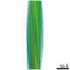
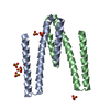
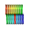
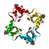
| タイトル | Cryo-EM structure of amyloid fibril formed by FUS low complexity domain | ||||||
 要素 要素 | fusion protein of mCerulean and FUS LCD | ||||||
 キーワード キーワード | PROTEIN FIBRIL / amyloid fibril | ||||||
| 機能・相同性 |  機能・相同性情報 機能・相同性情報mRNA stabilization / intracellular membraneless organelle / regulation of RNA splicing / Processing of Capped Intron-Containing Pre-mRNA / positive regulation of double-strand break repair via homologous recombination / mRNA Splicing - Major Pathway / RNA splicing / mRNA 3'-UTR binding / molecular condensate scaffold activity / transcription coregulator activity ...mRNA stabilization / intracellular membraneless organelle / regulation of RNA splicing / Processing of Capped Intron-Containing Pre-mRNA / positive regulation of double-strand break repair via homologous recombination / mRNA Splicing - Major Pathway / RNA splicing / mRNA 3'-UTR binding / molecular condensate scaffold activity / transcription coregulator activity / protein homooligomerization / amyloid fibril formation / transcription coactivator activity / chromatin binding / regulation of DNA-templated transcription / regulation of transcription by RNA polymerase II / DNA binding / RNA binding / nucleoplasm / identical protein binding / nucleus / metal ion binding / cytoplasm 類似検索 - 分子機能 | ||||||
| 生物種 |   Aequorea victoria (オワンクラゲ) Aequorea victoria (オワンクラゲ) Homo sapiens (ヒト) Homo sapiens (ヒト) | ||||||
| 手法 | 電子顕微鏡法 / らせん対称体再構成法 / クライオ電子顕微鏡法 / 解像度: 2.9 Å | ||||||
 データ登録者 データ登録者 | Sun, Y.P. / Xia, W.C. / Liu, C. | ||||||
| 資金援助 | 1件
| ||||||
 引用 引用 | ジャーナル: iScience / 年: 2022 タイトル: Molecular structure of an amyloid fibril formed by FUS low-complexity domain. 著者: Yunpeng Sun / Shenqing Zhang / Jiaojiao Hu / Youqi Tao / Wencheng Xia / Jinge Gu / Yichen Li / Qin Cao / Dan Li / Cong Liu /  要旨: FUS is a multifunctional nuclear protein which undergoes liquid-liquid phase separation in response to stress and DNA damage. Dysregulation of FUS dynamic phase separation leads to formation of ...FUS is a multifunctional nuclear protein which undergoes liquid-liquid phase separation in response to stress and DNA damage. Dysregulation of FUS dynamic phase separation leads to formation of pathological fibril closely associated with neurodegenerative diseases such as amyotrophic lateral sclerosis and frontotemporal dementia. In this study, we determined the cryo-EM structure of a cytotoxic fibril formed by the low-complexity (LC) domain of FUS at 2.9 Å resolution. The fibril structure exhibits a new and extensive serpentine fold consisting of three motifs incorporating together via a Tyr triad. FUS LC employs 91 residues to form an enlarged and stable fibril core via hydrophilic interaction and hydrogen bonds, which is distinct from most of previously determined fibrils commonly stabilized by hydrophobic interaction. Our work reveals the structural basis underlying formation of a cytotoxic and thermostable fibril of FUS LC and sheds light on understanding the liquid-to-solid phase transition of FUS in disease. | ||||||
| 履歴 |
|
- 構造の表示
構造の表示
| ムービー |
ムービービューア |
|---|---|
| 構造ビューア | 分子: MolmilJmol/JSmol |
- ダウンロードとリンク
-ダウンロード
| PDBx/mmCIF形式 | 7vqq.cif.gz | 71.8 KB | 表示 | PDBx/mmCIF形式 |
|---|---|---|---|---|
| PDB形式 | pdb7vqq.ent.gz | 43.2 KB | 表示 | PDB形式 |
| PDBx/mmJSON形式 | 7vqq.json.gz | ツリー表示 | PDBx/mmJSON形式 | |
| その他 |  その他のダウンロード その他のダウンロード |
-検証レポート
| 文書・要旨 | 7vqq_validation.pdf.gz | 881.3 KB | 表示 | wwPDB検証レポート |
|---|---|---|---|---|
| 文書・詳細版 | 7vqq_full_validation.pdf.gz | 881.4 KB | 表示 | |
| XML形式データ | 7vqq_validation.xml.gz | 18.8 KB | 表示 | |
| CIF形式データ | 7vqq_validation.cif.gz | 27 KB | 表示 | |
| アーカイブディレクトリ | https://data.pdbj.org/pub/pdb/validation_reports/vq/7vqqftp://data.pdbj.org/pub/pdb/validation_reports/vq/7vqq | HTTPS FTP |
-関連構造データ
-リンク
 PDBj
PDBj


- 集合体
集合体
| 登録構造単位 | 
|
|---|---|
| 1 |
|
-要素
| #1: タンパク質 | 分子量: 51948.352 Da / 分子数: 3 / 由来タイプ: 組換発現 詳細: 6His-tagged mCerulean:(FPbase ID: J2JWA, Link: https://www.fpbase.org/protein/mcerulean/) 由来: (組換発現) Aequorea victoria (オワンクラゲ), (組換発現) Homo sapiens (ヒト)遺伝子: FUS, TLS / 発現宿主:  |
|---|
-実験情報
-実験
| 実験 | 手法: 電子顕微鏡法 |
|---|---|
| EM実験 | 試料の集合状態: FILAMENT / 3次元再構成法: らせん対称体再構成法 |
- 試料調製
試料調製
| 構成要素 | 名称: Amyloid fibril formed by FUS low complexity domain / タイプ: ORGANELLE OR CELLULAR COMPONENT / Entity ID: all / 由来: RECOMBINANT | ||||||||||||
|---|---|---|---|---|---|---|---|---|---|---|---|---|---|
| 分子量 | 実験値: NO | ||||||||||||
| 由来(天然) |
| ||||||||||||
| 由来(組換発現) |
| ||||||||||||
| 緩衝液 | pH: 7.5 | ||||||||||||
| 試料 | 包埋: NO / シャドウイング: NO / 染色: NO / 凍結: YES | ||||||||||||
| 急速凍結 | 凍結剤: ETHANE |
- 電子顕微鏡撮影
| 実験機器 |  モデル: Titan Krios / 画像提供: FEI Company |
|---|---|
| 顕微鏡 | モデル: FEI TITAN KRIOS |
| 電子銃 | 電子線源:  FIELD EMISSION GUN / 加速電圧: 300 kV / 照射モード: FLOOD BEAM FIELD EMISSION GUN / 加速電圧: 300 kV / 照射モード: FLOOD BEAM |
| 電子レンズ | モード: BRIGHT FIELD |
| 撮影 | 電子線照射量: 40 e/Å2 フィルム・検出器のモデル: GATAN K2 SUMMIT (4k x 4k) |
- 解析
解析
| CTF補正 | タイプ: NONE |
|---|---|
| らせん対称 | 回転角度/サブユニット: -1.44 ° / 軸方向距離/サブユニット: 5 Å / らせん対称軸の対称性: C1 |
| 3次元再構成 | 解像度: 2.9 Å / 解像度の算出法: FSC 0.143 CUT-OFF / 粒子像の数: 171163 / 対称性のタイプ: HELICAL |
