 Movie
Movie Controller

[English] 日本語
 Yorodumi
- PDB-7vqc: Structure of MA1831 from Methanosarcina acetivorans in complex wi... -
+ Open data

- Basic information
| Entry | Database: PDB / ID: 7vqc | ||||||
|---|---|---|---|---|---|---|---|
| Title | Structure of MA1831 from Methanosarcina acetivorans in complex with pyrophosphate | ||||||
 Components Components | Di-trans-poly-cis-decaprenylcistransferase | ||||||
 Keywords Keywords | TRANSFERASE / cis-prenyltransferase | ||||||
| Function / homology |  Function and homology information Function and homology informationpolyprenol biosynthetic process / transferase activity, transferring alkyl or aryl (other than methyl) groups / metal ion binding Similarity search - Function | ||||||
| Biological species |  Methanosarcina acetivorans (archaea) Methanosarcina acetivorans (archaea) | ||||||
| Method |  X-RAY DIFFRACTION / MOLECULAR REPLACEMENT / Resolution: 2.45 Å X-RAY DIFFRACTION / MOLECULAR REPLACEMENT / Resolution: 2.45 Å | ||||||
 Authors Authors | Zhang, L.L. / Chen, C.C. / Liu, W.D. / Huang, J.W. / Zhang, X.W. / Liu, B.B. / Guo, R.T. | ||||||
| Funding support | 1items
| ||||||
 Citation Citation | Journal: Int.J.Biol.Macromol. / Year: 2022 Title: Structural insights to a bi-functional isoprenyl diphosphate synthase that can catalyze head-to-tail and head-to-middle condensation. Authors: Zhang, L. / Zhang, X. / Min, J. / Liu, B. / Huang, J.W. / Yang, Y. / Liu, W. / Dai, L. / Yang, Y. / Chen, C.C. / Guo, R.T. | ||||||
| History |
|
- Structure visualization
| Structure viewer | Molecule: MolmilJmol/JSmol |
|---|
- Downloads & links
-Download
| PDBx/mmCIF format | 7vqc.cif.gz | 102.2 KB | Display | PDBx/mmCIF format |
|---|---|---|---|---|
| PDB format | pdb7vqc.ent.gz | 76.9 KB | Display | PDB format |
| PDBx/mmJSON format | 7vqc.json.gz | Tree view | PDBx/mmJSON format | |
| Others |  Other downloads Other downloads |
-Validation report
| Arichive directory | https://data.pdbj.org/pub/pdb/validation_reports/vq/7vqcftp://data.pdbj.org/pub/pdb/validation_reports/vq/7vqc | HTTPS FTP |
|---|
-Related structure data
| Related structure data |  7vq9C  7vqaC  7vqbC  7vqdC  7caqS S: Starting model for refinement C: citing same article ( |
|---|---|
| Similar structure data |
-Links
 PDBj
PDBj


- Assembly
| Deposited unit | 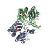
| ||||||||
|---|---|---|---|---|---|---|---|---|---|
| 1 |
| ||||||||
| Unit cell |
|
-Components
| #1: Protein | Mass: 25824.670 Da / Num. of mol.: 2 Source method: isolated from a genetically manipulated source Source: (gene. exp.) Methanosarcina acetivorans (strain ATCC 35395 / DSM 2834 / JCM 12185 / C2A) (archaea)Gene: uppS, MA_1831 / Plasmid: pET32a / Production host:  #2: Chemical |   Mass: 96.063 Da / Num. of mol.: 2 / Source method: obtained synthetically / Formula: SO4 Mass: 96.063 Da / Num. of mol.: 2 / Source method: obtained synthetically / Formula: SO4#3: Chemical | ChemComp-POP / |   Mass: 175.959 Da / Num. of mol.: 1 / Source method: obtained synthetically / Formula: H2O7P2 / Feature type: SUBJECT OF INVESTIGATION Mass: 175.959 Da / Num. of mol.: 1 / Source method: obtained synthetically / Formula: H2O7P2 / Feature type: SUBJECT OF INVESTIGATION#4: Water | ChemComp-HOH / |  Mass: 18.015 Da / Num. of mol.: 264 / Source method: isolated from a natural source / Formula: H2O Mass: 18.015 Da / Num. of mol.: 264 / Source method: isolated from a natural source / Formula: H2OHas ligand of interest | Y | |
|---|
-Experimental details
-Experiment
| Experiment | Method: X-RAY DIFFRACTION / Number of used crystals: 1 |
|---|
- Sample preparation
| Crystal | Density Matthews: 2.57 Å3/Da / Density % sol: 52.18 % |
|---|---|
| Crystal grow | Temperature: 298 K / Method: vapor diffusion, sitting drop / pH: 8 Details: 1.5M ammonium sulfate, 0.1M Tris (pH 8.0), 12% glycerol |
-Data collection
| Diffraction | Mean temperature: 100 K / Serial crystal experiment: N |
|---|---|
| Diffraction source | Source: LIQUID ANODE / Type: BRUKER METALJET / Wavelength: 1.34138 Å |
| Detector | Type: Bruker PHOTON III / Detector: PIXEL / Date: Apr 14, 2021 |
| Radiation | Protocol: SINGLE WAVELENGTH / Monochromatic (M) / Laue (L): M / Scattering type: x-ray |
| Radiation wavelength | Wavelength: 1.34138 Å / Relative weight: 1 |
| Reflection | Resolution: 2.29→33 Å / Num. obs: 19356 / % possible obs: 99 % / Redundancy: 6.2 % / Rmerge(I) obs: 0.188 / Net I/σ(I): 7.9 |
| Reflection shell | Resolution: 2.29→2.37 Å / Redundancy: 4.1 % / Rmerge(I) obs: 0.831 / Num. unique obs: 2085 / % possible all: 90.7 |
- Processing
| Software |
| ||||||||||||||||||||||||
|---|---|---|---|---|---|---|---|---|---|---|---|---|---|---|---|---|---|---|---|---|---|---|---|---|---|
| Refinement | Method to determine structure: MOLECULAR REPLACEMENT Starting model: 7CAQ Resolution: 2.45→33 Å / Cor.coef. Fo:Fc: 0.952 / Cor.coef. Fo:Fc free: 0.884 / SU B: 10.17 / SU ML: 0.223 / Cross valid method: THROUGHOUT / σ(F): 0 / ESU R: 0.481 / ESU R Free: 0.29 / Stereochemistry target values: MAXIMUM LIKELIHOOD Details: HYDROGENS HAVE BEEN ADDED IN THE RIDING POSITIONS U VALUES : REFINED INDIVIDUALLY
| ||||||||||||||||||||||||
| Solvent computation | Ion probe radii: 0.8 Å / Shrinkage radii: 0.8 Å / VDW probe radii: 1.2 Å / Solvent model: MASK | ||||||||||||||||||||||||
| Displacement parameters | Biso max: 119.09 Å2 / Biso mean: 30.67 Å2 / Biso min: 10.73 Å2
| ||||||||||||||||||||||||
| Refinement step | Cycle: final / Resolution: 2.45→33 Å
| ||||||||||||||||||||||||
| LS refinement shell | Resolution: 2.45→2.51 Å / Rfactor Rfree error: 0 / Total num. of bins used: 20
|
