 Movie
Movie Controller

[English] 日本語
 Yorodumi
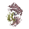
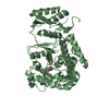
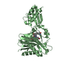
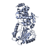
- PDB-7vnb: Crystal structure of the SARS-CoV-2 RBD in complex with a human s... -
+ Open data

- Basic information
| Entry | Database: PDB / ID: 7vnb | ||||||
|---|---|---|---|---|---|---|---|
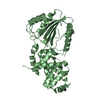
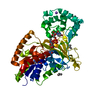
| Title | Crystal structure of the SARS-CoV-2 RBD in complex with a human single domain antibody n3113 | ||||||
 Components Components |
| ||||||
 Keywords Keywords | VIRAL PROTEIN / SARS-CoV-2 / nanobody | ||||||
| Function / homology |  Function and homology information Function and homology informationsymbiont-mediated disruption of host tissue / Maturation of spike protein / Translation of Structural Proteins / Virion Assembly and Release / host cell surface / host extracellular region / symbiont-mediated-mediated suppression of host tetherin activity / Induction of Cell-Cell Fusion / structural constituent of virion / positive regulation of viral entry into host cell ...symbiont-mediated disruption of host tissue / Maturation of spike protein / Translation of Structural Proteins / Virion Assembly and Release / host cell surface / host extracellular region / symbiont-mediated-mediated suppression of host tetherin activity / Induction of Cell-Cell Fusion / structural constituent of virion / positive regulation of viral entry into host cell / membrane fusion / host cell endoplasmic reticulum-Golgi intermediate compartment membrane / Attachment and Entry / entry receptor-mediated virion attachment to host cell / receptor-mediated virion attachment to host cell / host cell surface receptor binding / symbiont-mediated suppression of host innate immune response / endocytosis involved in viral entry into host cell / receptor ligand activity / fusion of virus membrane with host plasma membrane / fusion of virus membrane with host endosome membrane / viral envelope / symbiont entry into host cell / virion attachment to host cell / host cell plasma membrane / SARS-CoV-2 activates/modulates innate and adaptive immune responses / virion membrane / membrane / identical protein binding / plasma membrane Similarity search - Function | ||||||
| Biological species |  Homo sapiens (human) Homo sapiens (human)  Severe acute respiratory syndrome coronavirus 2 Severe acute respiratory syndrome coronavirus 2 | ||||||
| Method |  X-RAY DIFFRACTION / SYNCHROTRON / MOLECULAR REPLACEMENT / Resolution: 2.27 Å X-RAY DIFFRACTION / SYNCHROTRON / MOLECULAR REPLACEMENT / Resolution: 2.27 Å | ||||||
 Authors Authors | Yang, Z. / Wang, Y. / Kong, Y. / Jin, Y. / Wu, Y. / Ying, T. | ||||||
| Funding support |  China, 1items China, 1items
| ||||||
 Citation Citation | Journal: Signal Transduct Target Ther / Year: 2021 Title: A non-ACE2 competing human single-domain antibody confers broad neutralization against SARS-CoV-2 and circulating variants. Authors: Zhenlin Yang / Yulu Wang / Yujia Jin / Yuanfei Zhu / Yanling Wu / Cheng Li / Yu Kong / Wenping Song / Xiaolong Tian / Wuqiang Zhan / Ailing Huang / Shanshan Zhou / Shuai Xia / Xiaoxu Tian / ...Authors: Zhenlin Yang / Yulu Wang / Yujia Jin / Yuanfei Zhu / Yanling Wu / Cheng Li / Yu Kong / Wenping Song / Xiaolong Tian / Wuqiang Zhan / Ailing Huang / Shanshan Zhou / Shuai Xia / Xiaoxu Tian / Chao Peng / Cuicui Chen / Yibing Shi / Gaowei Hu / Shujuan Du / Yuyan Wang / Youhua Xie / Shibo Jiang / Lu Lu / Lei Sun / Yuanlin Song / Tianlei Ying / Abstract: The current COVID-19 pandemic has heavily burdened the global public health system and may keep simmering for years. The frequent emergence of immune escape variants have spurred the search for ...The current COVID-19 pandemic has heavily burdened the global public health system and may keep simmering for years. The frequent emergence of immune escape variants have spurred the search for prophylactic vaccines and therapeutic antibodies that confer broad protection against SARS-CoV-2 variants. Here we show that the bivalency of an affinity maturated fully human single-domain antibody (n3113.1-Fc) exhibits exquisite neutralizing potency against SARS-CoV-2 pseudovirus, and confers effective prophylactic and therapeutic protection against authentic SARS-CoV-2 in the host cell receptor angiotensin-converting enzyme 2 (ACE2) humanized mice. The crystal structure of n3113 in complex with the receptor-binding domain (RBD) of SARS-CoV-2, combined with the cryo-EM structures of n3113 and spike ecto-domain, reveals that n3113 binds to the side surface of up-state RBD with no competition with ACE2. The binding of n3113 to this novel epitope stabilizes spike in up-state conformations but inhibits SARS-CoV-2 S mediated membrane fusion, expanding our recognition of neutralization by antibodies against SARS-CoV-2. Binding assay and pseudovirus neutralization assay show no evasion of recently prevalent SARS-CoV-2 lineages, including Alpha (B.1.1.7), Beta (B.1.351), Gamma (P.1), and Delta (B.1.617.2) for n3113.1-Fc with Y58L mutation, demonstrating the potential of n3113.1-Fc (Y58L) as a promising candidate for clinical development to treat COVID-19. | ||||||
| History |
|
- Structure visualization
| Structure viewer | Molecule: MolmilJmol/JSmol |
|---|
- Downloads & links
-Download
| PDBx/mmCIF format | 7vnb.cif.gz | 103.9 KB | Display | PDBx/mmCIF format |
|---|---|---|---|---|
| PDB format | pdb7vnb.ent.gz | 63.6 KB | Display | PDB format |
| PDBx/mmJSON format | 7vnb.json.gz | Tree view | PDBx/mmJSON format | |
| Others |  Other downloads Other downloads |
-Validation report
| Arichive directory | https://data.pdbj.org/pub/pdb/validation_reports/vn/7vnbftp://data.pdbj.org/pub/pdb/validation_reports/vn/7vnb | HTTPS FTP |
|---|
-Related structure data
| Related structure data |  7vncC  7vndC  7vneC 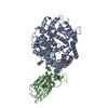 6lzgS S: Starting model for refinement C: citing same article ( |
|---|---|
| Similar structure data |


-Links
 PDBj
PDBj


- Assembly
| Deposited unit | 
| ||||||||||||
|---|---|---|---|---|---|---|---|---|---|---|---|---|---|
| 1 |
| ||||||||||||
| Unit cell |
|
-Components
| #1: Protein | Mass: 12963.374 Da / Num. of mol.: 1 Source method: isolated from a genetically manipulated source Source: (gene. exp.) Homo sapiens (human) / Production host:  |
|---|---|
| #2: Protein | Mass: 24966.086 Da / Num. of mol.: 1 Source method: isolated from a genetically manipulated source Source: (gene. exp.) Severe acute respiratory syndrome coronavirus 2Gene: S, 2 / Production host:   Spodoptera frugiperda (fall armyworm) / References: UniProt: P0DTC2 Spodoptera frugiperda (fall armyworm) / References: UniProt: P0DTC2 |
| #3: Sugar | ChemComp-NAG /   Type: D-saccharide, beta linking / Mass: 221.208 Da / Num. of mol.: 1 / Source method: obtained synthetically / Formula: C8H15NO6 Type: D-saccharide, beta linking / Mass: 221.208 Da / Num. of mol.: 1 / Source method: obtained synthetically / Formula: C8H15NO6 |
| #4: Water | ChemComp-HOH /  Mass: 18.015 Da / Num. of mol.: 245 / Source method: isolated from a natural source / Formula: H2O Mass: 18.015 Da / Num. of mol.: 245 / Source method: isolated from a natural source / Formula: H2O |
| Has ligand of interest | N |
| Has protein modification | Y |
-Experimental details
-Experiment
| Experiment | Method: X-RAY DIFFRACTION / Number of used crystals: 1 |
|---|
- Sample preparation
| Crystal | Density Matthews: 5.05 Å3/Da / Density % sol: 75.63 % |
|---|---|
| Crystal grow | Temperature: 289 K / Method: vapor diffusion, sitting drop / Details: 4.3M sodium chloride and 0.1M HEPES, pH 7.5 |
-Data collection
| Diffraction | Mean temperature: 100 K / Serial crystal experiment: N |
|---|---|
| Diffraction source | Source: SYNCHROTRON / Site: SSRF / Beamline: BL17U1 / Wavelength: 0.979 Å |
| Detector | Type: DECTRIS PILATUS 6M / Detector: PIXEL / Date: Jun 7, 2020 |
| Radiation | Protocol: SINGLE WAVELENGTH / Monochromatic (M) / Laue (L): M / Scattering type: x-ray |
| Radiation wavelength | Wavelength: 0.979 Å / Relative weight: 1 |
| Reflection | Resolution: 2.27→73.11 Å / Num. obs: 34264 / % possible obs: 99.9 % / Redundancy: 10.4 % / Biso Wilson estimate: 37.75 Å2 / CC1/2: 0.997 / Net I/σ(I): 11.1 |
| Reflection shell | Resolution: 2.27→2.33 Å / Num. unique obs: 2535 / CC1/2: 0.798 |
- Processing
| Software |
| |||||||||||||||||||||||||||||||||||||||||||||||||||||||||||||||||||||||||||||||||||||||||||||||||||||||||
|---|---|---|---|---|---|---|---|---|---|---|---|---|---|---|---|---|---|---|---|---|---|---|---|---|---|---|---|---|---|---|---|---|---|---|---|---|---|---|---|---|---|---|---|---|---|---|---|---|---|---|---|---|---|---|---|---|---|---|---|---|---|---|---|---|---|---|---|---|---|---|---|---|---|---|---|---|---|---|---|---|---|---|---|---|---|---|---|---|---|---|---|---|---|---|---|---|---|---|---|---|---|---|---|---|---|---|
| Refinement | Method to determine structure: MOLECULAR REPLACEMENT Starting model: 6LZG Resolution: 2.27→42.56 Å / SU ML: 0.2658 / Cross valid method: FREE R-VALUE / σ(F): 1.97 / Phase error: 23.9074
| |||||||||||||||||||||||||||||||||||||||||||||||||||||||||||||||||||||||||||||||||||||||||||||||||||||||||
| Solvent computation | Shrinkage radii: 0.9 Å / VDW probe radii: 1.11 Å / Solvent model: FLAT BULK SOLVENT MODEL | |||||||||||||||||||||||||||||||||||||||||||||||||||||||||||||||||||||||||||||||||||||||||||||||||||||||||
| Displacement parameters | Biso mean: 40.35 Å2 | |||||||||||||||||||||||||||||||||||||||||||||||||||||||||||||||||||||||||||||||||||||||||||||||||||||||||
| Refinement step | Cycle: LAST / Resolution: 2.27→42.56 Å
| |||||||||||||||||||||||||||||||||||||||||||||||||||||||||||||||||||||||||||||||||||||||||||||||||||||||||
| Refine LS restraints |
| |||||||||||||||||||||||||||||||||||||||||||||||||||||||||||||||||||||||||||||||||||||||||||||||||||||||||
| LS refinement shell |
|
