 Movie
Movie Controller


+ Open data

- Basic information
| Entry | Database: PDB / ID: 7vm8 | ||||||
|---|---|---|---|---|---|---|---|
| Title | Crystal structure of the MtDMI1 gating ring | ||||||
 Components Components | Ion channel DMI1 | ||||||
 Keywords Keywords | PLANT PROTEIN / DMI / symbiose | ||||||
| Function / homology | CASTOR/POLLUX/SYM8 ion channel, conserved domain / Ion channel CASTOR/POLLUX/SYM8-like / Castor and Pollux, part of voltage-gated ion channel / Calcium-activated potassium channel slowpoke-like RCK domain / RCK N-terminal domain profile. / nuclear membrane / monoatomic ion transmembrane transport / NAD(P)-binding domain superfamily / Ion channel DMI1 Function and homology information Function and homology information | ||||||
| Biological species |  | ||||||
| Method |  X-RAY DIFFRACTION / SYNCHROTRON / MOLECULAR REPLACEMENT / Resolution: 3.034 Å X-RAY DIFFRACTION / SYNCHROTRON / MOLECULAR REPLACEMENT / Resolution: 3.034 Å | ||||||
 Authors Authors | Huang, X. / Zhang, P. | ||||||
| Funding support | 1items
| ||||||
 Citation Citation | Journal: Proc.Natl.Acad.Sci.USA / Year: 2022 Title: Constitutive activation of a nuclear-localized calcium channel complex in Medicago truncatula. Authors: Liu, H. / Lin, J.S. / Luo, Z. / Sun, J. / Huang, X. / Yang, Y. / Xu, J. / Wang, Y.F. / Zhang, P. / Oldroyd, G.E.D. / Xie, F. | ||||||
| History |
|
- Structure visualization
| Structure viewer | Molecule: MolmilJmol/JSmol |
|---|
- Downloads & links
-Download
| PDBx/mmCIF format | 7vm8.cif.gz | 112.7 KB | Display | PDBx/mmCIF format |
|---|---|---|---|---|
| PDB format | pdb7vm8.ent.gz | 84.4 KB | Display | PDB format |
| PDBx/mmJSON format | 7vm8.json.gz | Tree view | PDBx/mmJSON format | |
| Others |  Other downloads Other downloads |
-Validation report
| Arichive directory | https://data.pdbj.org/pub/pdb/validation_reports/vm/7vm8ftp://data.pdbj.org/pub/pdb/validation_reports/vm/7vm8 | HTTPS FTP |
|---|
-Related structure data
| Related structure data | 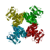 6o6jS S: Starting model for refinement |
|---|---|
| Similar structure data |
-Links
 PDBj
PDBj
- Assembly
| Deposited unit | 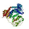
| ||||||||
|---|---|---|---|---|---|---|---|---|---|
| 1 |
| ||||||||
| Unit cell |
|
-Components
| #1: Protein | Mass: 60475.391 Da / Num. of mol.: 1 Source method: isolated from a genetically manipulated source Source: (gene. exp.)  Komagataella pastoris (fungus) / Strain (production host): GS115 / References: UniProt: Q6RHR6 Komagataella pastoris (fungus) / Strain (production host): GS115 / References: UniProt: Q6RHR6 |
|---|
-Experimental details
-Experiment
| Experiment | Method: X-RAY DIFFRACTION / Number of used crystals: 1 |
|---|
- Sample preparation
| Crystal | Density Matthews: 2.33 Å3/Da / Density % sol: 47.14 % |
|---|---|
| Crystal grow | Temperature: 293 K / Method: vapor diffusion, sitting drop / pH: 5.4 Details: 16% (wt/vol) PEG 4000, 0.2M KCl, 0.01M CaCl2, 0.05 M Sodium cacodylate trihydrate pH 5.4 |
-Data collection
| Diffraction | Mean temperature: 100 K / Serial crystal experiment: N | |||||||||||||||||||||||||||||||||||||||||||||||||||||||||||||||||||||||||||||||||||||||||||||||||||
|---|---|---|---|---|---|---|---|---|---|---|---|---|---|---|---|---|---|---|---|---|---|---|---|---|---|---|---|---|---|---|---|---|---|---|---|---|---|---|---|---|---|---|---|---|---|---|---|---|---|---|---|---|---|---|---|---|---|---|---|---|---|---|---|---|---|---|---|---|---|---|---|---|---|---|---|---|---|---|---|---|---|---|---|---|---|---|---|---|---|---|---|---|---|---|---|---|---|---|---|---|
| Diffraction source | Source: SYNCHROTRON / Site: SSRF  / Beamline: BL19U1 / Wavelength: 0.9793 Å / Beamline: BL19U1 / Wavelength: 0.9793 Å | |||||||||||||||||||||||||||||||||||||||||||||||||||||||||||||||||||||||||||||||||||||||||||||||||||
| Detector | Type: MAR555 FLAT PANEL / Detector: IMAGE PLATE / Date: Oct 10, 2019 | |||||||||||||||||||||||||||||||||||||||||||||||||||||||||||||||||||||||||||||||||||||||||||||||||||
| Radiation | Protocol: SINGLE WAVELENGTH / Monochromatic (M) / Laue (L): M / Scattering type: x-ray | |||||||||||||||||||||||||||||||||||||||||||||||||||||||||||||||||||||||||||||||||||||||||||||||||||
| Radiation wavelength | Wavelength: 0.9793 Å / Relative weight: 1 | |||||||||||||||||||||||||||||||||||||||||||||||||||||||||||||||||||||||||||||||||||||||||||||||||||
| Reflection | Resolution: 3→50 Å / Num. obs: 14703 / % possible obs: 100 % / Redundancy: 13.1 % / Rmerge(I) obs: 0.087 / Rpim(I) all: 0.025 / Rrim(I) all: 0.09 / Χ2: 0.94 / Net I/σ(I): 7.1 | |||||||||||||||||||||||||||||||||||||||||||||||||||||||||||||||||||||||||||||||||||||||||||||||||||
| Reflection shell | Diffraction-ID: 1
|
- Processing
| Software |
| ||||||||||||||||||||||||||||||||||||
|---|---|---|---|---|---|---|---|---|---|---|---|---|---|---|---|---|---|---|---|---|---|---|---|---|---|---|---|---|---|---|---|---|---|---|---|---|---|
| Refinement | Method to determine structure: MOLECULAR REPLACEMENT Starting model: 6O6J Resolution: 3.034→45.8 Å / SU ML: 0.48 / Cross valid method: THROUGHOUT / σ(F): 1.34 / Phase error: 36.53 / Stereochemistry target values: ML
| ||||||||||||||||||||||||||||||||||||
| Solvent computation | Shrinkage radii: 0.9 Å / VDW probe radii: 1.11 Å / Solvent model: FLAT BULK SOLVENT MODEL | ||||||||||||||||||||||||||||||||||||
| Displacement parameters | Biso max: 138.14 Å2 / Biso mean: 47.0991 Å2 / Biso min: 14.64 Å2 | ||||||||||||||||||||||||||||||||||||
| Refinement step | Cycle: final / Resolution: 3.034→45.8 Å
| ||||||||||||||||||||||||||||||||||||
| LS refinement shell | Refine-ID: X-RAY DIFFRACTION / Rfactor Rfree error: 0
|
