 Movie
Movie Controller


+ Open data

- Basic information
| Entry | Database: PDB / ID: 7vla | ||||||
|---|---|---|---|---|---|---|---|
| Title | Cryo-EM structure of the CCL15(27-92) bound CCR1-Gi complex | ||||||
 Components Components |
| ||||||
 Keywords Keywords | MEMBRANE PROTEIN / GPCR / CCR1 / Chemokine Receptor / MEMBRNE PROTEIN | ||||||
| Function / homology |  Function and homology information Function and homology informationchemokine (C-C motif) ligand 7 binding / chemokine (C-C motif) ligand 5 binding / chemokine receptor activity / CCR chemokine receptor binding / phosphatidylinositol-4,5-bisphosphate phospholipase C activity / negative regulation of bone mineralization / C-C chemokine binding / C-C chemokine receptor activity / positive regulation of calcium ion transport / positive regulation of osteoclast differentiation ...chemokine (C-C motif) ligand 7 binding / chemokine (C-C motif) ligand 5 binding / chemokine receptor activity / CCR chemokine receptor binding / phosphatidylinositol-4,5-bisphosphate phospholipase C activity / negative regulation of bone mineralization / C-C chemokine binding / C-C chemokine receptor activity / positive regulation of calcium ion transport / positive regulation of osteoclast differentiation / positive regulation of monocyte chemotaxis / chemokine activity / Chemokine receptors bind chemokines / positive regulation of neuroinflammatory response / dendritic cell chemotaxis / chemoattractant activity / Interleukin-10 signaling / exocytosis / monocyte chemotaxis / G protein-coupled receptor signaling pathway, coupled to cyclic nucleotide second messenger / adenylate cyclase inhibitor activity / positive regulation of protein localization to cell cortex / T cell migration / positive regulation of relaxation of smooth muscle / Adenylate cyclase inhibitory pathway / D2 dopamine receptor binding / adenylate cyclase-inhibiting serotonin receptor signaling pathway / G protein-coupled serotonin receptor binding / cellular response to forskolin / regulation of mitotic spindle organization / chemokine-mediated signaling pathway / cell chemotaxis / calcium-mediated signaling / Regulation of insulin secretion / neuropeptide signaling pathway / response to prostaglandin E / positive regulation of cholesterol biosynthetic process / response to wounding / negative regulation of insulin secretion / G protein-coupled receptor binding / response to peptide hormone / chemotaxis / intracellular calcium ion homeostasis / centriolar satellite / cytokine-mediated signaling pathway / G-protein beta/gamma-subunit complex binding / adenylate cyclase-modulating G protein-coupled receptor signaling pathway / adenylate cyclase-inhibiting G protein-coupled receptor signaling pathway / Olfactory Signaling Pathway / positive regulation of inflammatory response / Activation of the phototransduction cascade / G protein-coupled acetylcholine receptor signaling pathway / G beta:gamma signalling through PLC beta / Presynaptic function of Kainate receptors / Thromboxane signalling through TP receptor / Activation of G protein gated Potassium channels / Inhibition of voltage gated Ca2+ channels via Gbeta/gamma subunits / G-protein activation / calcium ion transport / Glucagon signaling in metabolic regulation / G beta:gamma signalling through CDC42 / Prostacyclin signalling through prostacyclin receptor / Synthesis, secretion, and inactivation of Glucagon-like Peptide-1 (GLP-1) / G beta:gamma signalling through BTK / photoreceptor disc membrane / ADP signalling through P2Y purinoceptor 12 / GDP binding / Glucagon-type ligand receptors / Sensory perception of sweet, bitter, and umami (glutamate) taste / Adrenaline,noradrenaline inhibits insulin secretion / Vasopressin regulates renal water homeostasis via Aquaporins / Glucagon-like Peptide-1 (GLP1) regulates insulin secretion / G alpha (z) signalling events / cell-cell signaling / cellular response to catecholamine stimulus / ADP signalling through P2Y purinoceptor 1 / antimicrobial humoral immune response mediated by antimicrobial peptide / G beta:gamma signalling through PI3Kgamma / ADORA2B mediated anti-inflammatory cytokines production / heparin binding / adenylate cyclase-activating dopamine receptor signaling pathway / Cooperation of PDCL (PhLP1) and TRiC/CCT in G-protein beta folding / GPER1 signaling / cellular response to prostaglandin E stimulus / heterotrimeric G-protein complex / G alpha (12/13) signalling events / Inactivation, recovery and regulation of the phototransduction cascade / G-protein beta-subunit binding / extracellular vesicle / sensory perception of taste / Thrombin signalling through proteinase activated receptors (PARs) / sperm principal piece / adenylate cyclase-activating G protein-coupled receptor signaling pathway / signaling receptor complex adaptor activity / positive regulation of cytosolic calcium ion concentration / retina development in camera-type eye / fibroblast proliferation / GTPase binding / G protein activity / midbody Similarity search - Function | ||||||
| Biological species |  Homo sapiens (human) Homo sapiens (human) | ||||||
| Method | ELECTRON MICROSCOPY / single particle reconstruction / cryo EM / Resolution: 2.7 Å | ||||||
 Authors Authors | Shao, Z. / Shen, Q. / Mao, C. / Yao, B. / Chen, L. / Zhang, H. / Shen, D. / Zhang, C. / Li, W. / Du, X. ...Shao, Z. / Shen, Q. / Mao, C. / Yao, B. / Chen, L. / Zhang, H. / Shen, D. / Zhang, C. / Li, W. / Du, X. / Li, F. / Ma, H. / Chen, Z. / Xu, H.E. / Ying, S. / Zhang, Y. / Shen, H. | ||||||
| Funding support |  China, 1items China, 1items
| ||||||
 Citation Citation | Journal: Nat Chem Biol / Year: 2022 Title: Identification and mechanism of G protein-biased ligands for chemokine receptor CCR1. Authors: Zhehua Shao / Qingya Shen / Bingpeng Yao / Chunyou Mao / Li-Nan Chen / Huibing Zhang / Dan-Dan Shen / Chao Zhang / Weijie Li / Xufei Du / Fei Li / Honglei Ma / Zhi-Hua Chen / H Eric Xu / ...Authors: Zhehua Shao / Qingya Shen / Bingpeng Yao / Chunyou Mao / Li-Nan Chen / Huibing Zhang / Dan-Dan Shen / Chao Zhang / Weijie Li / Xufei Du / Fei Li / Honglei Ma / Zhi-Hua Chen / H Eric Xu / Songmin Ying / Yan Zhang / Huahao Shen / Abstract: Biased signaling of G protein-coupled receptors describes an ability of different ligands that preferentially activate an alternative downstream signaling pathway. In this work, we identified and ...Biased signaling of G protein-coupled receptors describes an ability of different ligands that preferentially activate an alternative downstream signaling pathway. In this work, we identified and characterized different N-terminal truncations of endogenous chemokine CCL15 as balanced or biased agonists targeting CCR1, and presented three cryogenic-electron microscopy structures of the CCR1-G complex in the ligand-free form or bound to different CCL15 truncations with a resolution of 2.6-2.9 Å, illustrating the structural basis of natural biased signaling that initiates an inflammation response. Complemented with pharmacological and computational studies, these structures revealed it was the conformational change of Tyr291 (Y291) in CCR1 that triggered its polar network rearrangement in the orthosteric binding pocket and allosterically regulated the activation of β-arrestin signaling. Our structure of CCL15-bound CCR1 also exhibited a critical site for ligand binding distinct from many other chemokine-receptor complexes, providing new insights into the mode of chemokine recognition. | ||||||
| History |
|
- Structure visualization
| Structure viewer | Molecule: MolmilJmol/JSmol |
|---|
- Downloads & links
-Download
| PDBx/mmCIF format | 7vla.cif.gz | 237 KB | Display | PDBx/mmCIF format |
|---|---|---|---|---|
| PDB format | pdb7vla.ent.gz | 177.3 KB | Display | PDB format |
| PDBx/mmJSON format | 7vla.json.gz | Tree view | PDBx/mmJSON format | |
| Others |  Other downloads Other downloads |
-Validation report
| Arichive directory | https://data.pdbj.org/pub/pdb/validation_reports/vl/7vlaftp://data.pdbj.org/pub/pdb/validation_reports/vl/7vla | HTTPS FTP |
|---|
-Related structure data
| Related structure data | 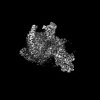 32022MC  7vl8C  7vl9C M: map data used to model this data C: citing same article ( |
|---|---|
| Similar structure data |


-Links
 PDBj
PDBj


- Assembly
| Deposited unit | 
|
|---|---|
| 1 |
|
-Components
-Guanine nucleotide-binding protein ... , 3 types, 3 molecules ABG
| #1: Protein | Mass: 40414.047 Da / Num. of mol.: 1 Source method: isolated from a genetically manipulated source Source: (gene. exp.) Homo sapiens (human) / Gene: GNAI1 / Cell (production host): SF9 / Production host:   Spodoptera frugiperda (fall armyworm) / References: UniProt: P63096 Spodoptera frugiperda (fall armyworm) / References: UniProt: P63096 |
|---|---|
| #2: Protein | Mass: 37728.152 Da / Num. of mol.: 1 Source method: isolated from a genetically manipulated source Source: (gene. exp.) Homo sapiens (human) / Gene: GNB1 / Production host: Spodoptera frugiperda (fall armyworm) / References: UniProt: P62873 |
| #3: Protein | Mass: 7861.143 Da / Num. of mol.: 1 Source method: isolated from a genetically manipulated source Source: (gene. exp.) Homo sapiens (human) / Gene: GNG2 / Cell (production host): SF9 / Production host: Spodoptera frugiperda (fall armyworm) / References: UniProt: P59768 |
-Protein , 2 types, 2 molecules LR
| #4: Protein | Mass: 8218.506 Da / Num. of mol.: 1 Source method: isolated from a genetically manipulated source Source: (gene. exp.) Homo sapiens (human) / Gene: CCL15, MIP5, NCC3, SCYA15 / Production host: Spodoptera frugiperda (fall armyworm) / References: UniProt: Q16663 |
|---|---|
| #5: Protein | Mass: 42203.547 Da / Num. of mol.: 1 Source method: isolated from a genetically manipulated source Source: (gene. exp.) Homo sapiens (human) / Gene: CCR1, CMKBR1, CMKR1, SCYAR1 / Production host: Spodoptera frugiperda (fall armyworm) / References: UniProt: P32246 |
-Antibody / Non-polymers , 2 types, 2 molecules S
| #6: Antibody | Mass: 27340.482 Da / Num. of mol.: 1 Source method: isolated from a genetically manipulated source Source: (gene. exp.) Homo sapiens (human) / Production host: Spodoptera frugiperda (fall armyworm) |
|---|---|
| #7: Chemical | ChemComp-CLR /  Mass: 386.654 Da / Num. of mol.: 1 / Source method: obtained synthetically / Formula: C27H46O / Feature type: SUBJECT OF INVESTIGATION Mass: 386.654 Da / Num. of mol.: 1 / Source method: obtained synthetically / Formula: C27H46O / Feature type: SUBJECT OF INVESTIGATION |
-Details
| Has ligand of interest | Y |
|---|---|
| Has protein modification | Y |
-Experimental details
-Experiment
| Experiment | Method: ELECTRON MICROSCOPY |
|---|---|
| EM experiment | Aggregation state: PARTICLE / 3D reconstruction method: single particle reconstruction |
- Sample preparation
| Component | Name: CCL15(27-92)-bound CCR1-Gi complex / Type: COMPLEX / Entity ID: #1-#6 / Source: RECOMBINANT |
|---|---|
| Source (natural) | Organism: Homo sapiens (human) |
| Source (recombinant) | Organism: Spodoptera frugiperda (fall armyworm) |
| Buffer solution | pH: 7.5 |
| Specimen | Embedding applied: NO / Shadowing applied: NO / Staining applied: NO / Vitrification applied: YES |
| Vitrification | Instrument: FEI VITROBOT MARK IV / Cryogen name: ETHANE |
- Electron microscopy imaging
| Experimental equipment |  Model: Titan Krios / Image courtesy: FEI Company |
|---|---|
| Microscopy | Model: FEI TITAN KRIOS |
| Electron gun | Electron source:  FIELD EMISSION GUN / Accelerating voltage: 300 kV / Illumination mode: FLOOD BEAM FIELD EMISSION GUN / Accelerating voltage: 300 kV / Illumination mode: FLOOD BEAM |
| Electron lens | Mode: BRIGHT FIELD |
| Image recording | Electron dose: 62 e/Å2 / Detector mode: COUNTING / Film or detector model: GATAN K2 SUMMIT (4k x 4k) |
- Processing
| Software |
| ||||||||||||||||||||||||
|---|---|---|---|---|---|---|---|---|---|---|---|---|---|---|---|---|---|---|---|---|---|---|---|---|---|
| EM software |
| ||||||||||||||||||||||||
| CTF correction | Type: PHASE FLIPPING AND AMPLITUDE CORRECTION | ||||||||||||||||||||||||
| 3D reconstruction | Resolution: 2.7 Å / Resolution method: FSC 0.143 CUT-OFF / Num. of particles: 423872 / Symmetry type: POINT | ||||||||||||||||||||||||
| Atomic model building | Protocol: FLEXIBLE FIT / Space: REAL | ||||||||||||||||||||||||
| Atomic model building | PDB-ID: 6DO1 Accession code: 6DO1 / Source name: PDB / Type: experimental model | ||||||||||||||||||||||||
| Refinement | Cross valid method: NONE Stereochemistry target values: GeoStd + Monomer Library + CDL v1.2 | ||||||||||||||||||||||||
| Displacement parameters | Biso mean: 39.9 Å2 | ||||||||||||||||||||||||
| Refine LS restraints |
|

