 Movie
Movie Controller

[English] 日本語
 Yorodumi
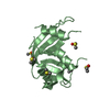
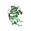
- PDB-7vjn: Crystal structure of anti-CRISPR-associated protein Aca1 in Pseud... -
+ Open data

- Basic information
| Entry | Database: PDB / ID: 7vjn | |||||||||
|---|---|---|---|---|---|---|---|---|---|---|
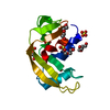
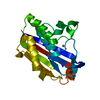
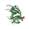
| Title | Crystal structure of anti-CRISPR-associated protein Aca1 in Pseudomonas phage JBD30 | |||||||||
 Components Components | anti-CRISPR-associated protein Aca1 | |||||||||
 Keywords Keywords | TRANSCRIPTION / DNA binding / autoregulation | |||||||||
| Function / homology | Lambda repressor-like, DNA-binding domain superfamily / DNA binding / Uncharacterized protein Function and homology information Function and homology information | |||||||||
| Biological species |  Pseudomonas phage JBD30 (virus) Pseudomonas phage JBD30 (virus) | |||||||||
| Method |  X-RAY DIFFRACTION / SYNCHROTRON / MOLECULAR REPLACEMENT / Resolution: 1.799 Å X-RAY DIFFRACTION / SYNCHROTRON / MOLECULAR REPLACEMENT / Resolution: 1.799 Å | |||||||||
 Authors Authors | Liu, Y.H. / Zhang, L.S. / Wu, B.X. / Huang, H.D. | |||||||||
| Funding support |  China, 1items China, 1items
| |||||||||
 Citation Citation | Journal: J.Biol.Chem. / Year: 2021 Title: Structural basis for anti-CRISPR repression mediated by bacterial operon proteins Aca1 and Aca2. Authors: Liu, Y. / Zhang, L. / Guo, M. / Chen, L. / Wu, B. / Huang, H. | |||||||||
| History |
|
- Structure visualization
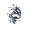
| Structure viewer | Molecule: MolmilJmol/JSmol |
|---|
- Downloads & links
-Download
| PDBx/mmCIF format | 7vjn.cif.gz | 73 KB | Display | PDBx/mmCIF format |
|---|---|---|---|---|
| PDB format | pdb7vjn.ent.gz | 52.9 KB | Display | PDB format |
| PDBx/mmJSON format | 7vjn.json.gz | Tree view | PDBx/mmJSON format | |
| Others |  Other downloads Other downloads |
-Validation report
| Arichive directory | https://data.pdbj.org/pub/pdb/validation_reports/vj/7vjnftp://data.pdbj.org/pub/pdb/validation_reports/vj/7vjn | HTTPS FTP |
|---|
-Related structure data
| Related structure data | 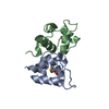 7fa3SC 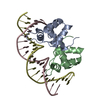 7vjmC 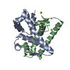 7vjoC 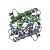 7vjpC 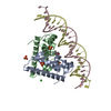 7vjqC S: Starting model for refinement C: citing same article ( |
|---|---|
| Similar structure data |


-Links
 PDBj
PDBj


- Assembly
| Deposited unit | 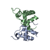
| ||||||||
|---|---|---|---|---|---|---|---|---|---|
| 1 |
| ||||||||
| Unit cell |
|
-Components
| #1: Protein | Mass: 9303.537 Da / Num. of mol.: 2 / Mutation: C71S Source method: isolated from a genetically manipulated source Source: (gene. exp.) Pseudomonas phage JBD30 (virus) / Gene: JBD30_036 / Production host:  #2: Water | ChemComp-HOH / |  Mass: 18.015 Da / Num. of mol.: 131 / Source method: isolated from a natural source / Formula: H2O Mass: 18.015 Da / Num. of mol.: 131 / Source method: isolated from a natural source / Formula: H2O |
|---|
-Experimental details
-Experiment
| Experiment | Method: X-RAY DIFFRACTION / Number of used crystals: 1 |
|---|
- Sample preparation
| Crystal | Density Matthews: 2.01 Å3/Da / Density % sol: 38.82 % |
|---|---|
| Crystal grow | Temperature: 293 K / Method: vapor diffusion Details: 0.2 M di-potassium hydrogen phosphate, 20% (w/v) PEG 3350 |
-Data collection
| Diffraction | Mean temperature: 100 K / Serial crystal experiment: N |
|---|---|
| Diffraction source | Source: SYNCHROTRON / Site: SSRF / Beamline: BL19U1 / Wavelength: 0.979 Å |
| Detector | Type: DECTRIS PILATUS3 S 6M / Detector: PIXEL / Date: Dec 29, 2019 |
| Radiation | Protocol: SINGLE WAVELENGTH / Monochromatic (M) / Laue (L): M / Scattering type: x-ray |
| Radiation wavelength | Wavelength: 0.979 Å / Relative weight: 1 |
| Reflection | Resolution: 1.799→40 Å / Num. obs: 14457 / % possible obs: 99.7 % / Redundancy: 10.2 % / Biso Wilson estimate: 27.7 Å2 / Rmerge(I) obs: 0.069 / Net I/σ(I): 29.4 |
| Reflection shell | Resolution: 1.8→1.86 Å / Num. unique obs: 1407 / CC1/2: 0.944 |
- Processing
| Software |
| ||||||||||||||||||||||||||||||||||||
|---|---|---|---|---|---|---|---|---|---|---|---|---|---|---|---|---|---|---|---|---|---|---|---|---|---|---|---|---|---|---|---|---|---|---|---|---|---|
| Refinement | Method to determine structure: MOLECULAR REPLACEMENT Starting model: 7FA3 Resolution: 1.799→39.829 Å / SU ML: 0.18 / Cross valid method: THROUGHOUT / σ(F): 1.38 / Phase error: 25.92 / Stereochemistry target values: ML
| ||||||||||||||||||||||||||||||||||||
| Solvent computation | Shrinkage radii: 0.9 Å / VDW probe radii: 1.11 Å / Solvent model: FLAT BULK SOLVENT MODEL | ||||||||||||||||||||||||||||||||||||
| Displacement parameters | Biso max: 94.99 Å2 / Biso mean: 37.2274 Å2 / Biso min: 18.14 Å2 | ||||||||||||||||||||||||||||||||||||
| Refinement step | Cycle: final / Resolution: 1.799→39.829 Å
| ||||||||||||||||||||||||||||||||||||
| Refine LS restraints |
| ||||||||||||||||||||||||||||||||||||
| LS refinement shell | Refine-ID: X-RAY DIFFRACTION / Rfactor Rfree error: 0
|
