 Movie
Movie Controller

[English] 日本語
 Yorodumi
- PDB-7vgg: Cryo-EM structure of Ultraviolet-B activated UVR8 in complex with COP1 -
+ Open data

- Basic information
| Entry | Database: PDB / ID: 7vgg | |||||||||||||||||||||||||||
|---|---|---|---|---|---|---|---|---|---|---|---|---|---|---|---|---|---|---|---|---|---|---|---|---|---|---|---|---|
| Title | Cryo-EM structure of Ultraviolet-B activated UVR8 in complex with COP1 | |||||||||||||||||||||||||||
 Components Components |
| |||||||||||||||||||||||||||
 Keywords Keywords | SIGNALING PROTEIN / COP1 / UVR8 | |||||||||||||||||||||||||||
| Function / homology |  Function and homology information Function and homology informationanthocyanin-containing compound metabolic process / shade avoidance / positive regulation of flavonoid biosynthetic process / skotomorphogenesis / photoperiodism, flowering / red, far-red light phototransduction / regulation of stomatal movement / photomorphogenesis / nuclear ubiquitin ligase complex / entrainment of circadian clock ...anthocyanin-containing compound metabolic process / shade avoidance / positive regulation of flavonoid biosynthetic process / skotomorphogenesis / photoperiodism, flowering / red, far-red light phototransduction / regulation of stomatal movement / photomorphogenesis / nuclear ubiquitin ligase complex / entrainment of circadian clock / response to UV-B / plastid / Cul4-RING E3 ubiquitin ligase complex / photoreceptor activity / response to UV / guanyl-nucleotide exchange factor activity / RING-type E3 ubiquitin transferase / ubiquitin-protein transferase activity / ubiquitin protein ligase activity / proteasome-mediated ubiquitin-dependent protein catabolic process / nuclear body / protein ubiquitination / DNA repair / chromatin binding / chromatin / protein homodimerization activity / zinc ion binding / identical protein binding / nucleus / cytoplasm / cytosol Similarity search - Function | |||||||||||||||||||||||||||
| Biological species |  | |||||||||||||||||||||||||||
| Method | ELECTRON MICROSCOPY / single particle reconstruction / cryo EM / Resolution: 3.1 Å | |||||||||||||||||||||||||||
 Authors Authors | Wang, Y.D. / Wang, L.X. / Guan, Z.Y. / Yin, P. | |||||||||||||||||||||||||||
| Funding support |  China, 1items China, 1items
| |||||||||||||||||||||||||||
 Citation Citation | Journal: Sci Adv / Year: 2022 Title: Structural insight into UV-B-activated UVR8 bound to COP1. Authors: Yidong Wang / Lixia Wang / Zeyuan Guan / Hongfei Chang / Ling Ma / Cuicui Shen / Liang Qiu / Junjie Yan / Delin Zhang / Jian Li / Xing Wang Deng / Ping Yin / Abstract: The CONSTITUTIVE PHOTOMORPHOGENIC 1-SUPPRESSOR OF PHYA-105 (COP1-SPA) complex is a central repressor of photomorphogenesis. This complex acts as an E3 ubiquitin ligase downstream of various light ...The CONSTITUTIVE PHOTOMORPHOGENIC 1-SUPPRESSOR OF PHYA-105 (COP1-SPA) complex is a central repressor of photomorphogenesis. This complex acts as an E3 ubiquitin ligase downstream of various light signaling transduced from multiple photoreceptors in plants. How the COP1-SPA activity is regulated by divergent light-signaling pathways remains largely elusive. Here, we reproduced the regulation pathway of COP1-SPA in ultraviolet-B (UV-B) signaling in vitro and determined the cryo-electron microscopy structure of UV-B receptor UVR8 in complex with COP1. The complex formation is mediated by two-interface interactions between UV-B-activated UVR8 and COP1. Both interfaces are essential for the competitive binding of UVR8 against the signaling hub component HY5 to the COP1-SPA complex. We also show that RUP2 dissociates UVR8 from the COP1-SPA4-UVR8 complex and facilitates its redimerization. Our results support a UV-B signaling model that the COP1-SPA activity is repressed by UV-B-activated UVR8 and derepressed by RUP2, owing to competitive binding, and provide a framework for studying the regulatory roles of distinct photoreceptors on photomorphogenesis. | |||||||||||||||||||||||||||
| History |
|
- Structure visualization
| Structure viewer | Molecule: MolmilJmol/JSmol |
|---|
- Downloads & links
-Download
| PDBx/mmCIF format | 7vgg.cif.gz | 140.3 KB | Display | PDBx/mmCIF format |
|---|---|---|---|---|
| PDB format | pdb7vgg.ent.gz | 99.8 KB | Display | PDB format |
| PDBx/mmJSON format | 7vgg.json.gz | Tree view | PDBx/mmJSON format | |
| Others |  Other downloads Other downloads |
-Validation report
| Arichive directory | https://data.pdbj.org/pub/pdb/validation_reports/vg/7vggftp://data.pdbj.org/pub/pdb/validation_reports/vg/7vgg | HTTPS FTP |
|---|
-Related structure data
| Related structure data |  31968MC M: map data used to model this data C: citing same article ( |
|---|---|
| Similar structure data |
-Links
 PDBj
PDBj

- Assembly
| Deposited unit | 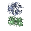
|
|---|---|
| 1 |
|
-Components
| #1: Protein | Mass: 79731.203 Da / Num. of mol.: 1 Source method: isolated from a genetically manipulated source Source: (gene. exp.)  Homo sapiens (human) Homo sapiens (human)References: UniProt: P43254, RING-type E3 ubiquitin transferase |
|---|---|
| #2: Protein | Mass: 50226.488 Da / Num. of mol.: 1 Source method: isolated from a genetically manipulated source Source: (gene. exp.)  |
| Has protein modification | N |
-Experimental details
-Experiment
| Experiment | Method: ELECTRON MICROSCOPY |
|---|---|
| EM experiment | Aggregation state: PARTICLE / 3D reconstruction method: single particle reconstruction |
- Sample preparation
| Component |
| ||||||||||||||||||||||||
|---|---|---|---|---|---|---|---|---|---|---|---|---|---|---|---|---|---|---|---|---|---|---|---|---|---|
| Source (natural) |
| ||||||||||||||||||||||||
| Source (recombinant) |
| ||||||||||||||||||||||||
| Buffer solution | pH: 7.5 | ||||||||||||||||||||||||
| Specimen | Conc.: 0.55 mg/ml / Embedding applied: NO / Shadowing applied: NO / Staining applied: NO / Vitrification applied: YES | ||||||||||||||||||||||||
| Vitrification | Cryogen name: ETHANE |
- Electron microscopy imaging
| Experimental equipment |  Model: Titan Krios / Image courtesy: FEI Company |
|---|---|
| Microscopy | Model: FEI TITAN KRIOS |
| Electron gun | Electron source:  FIELD EMISSION GUN / Accelerating voltage: 300 kV / Illumination mode: FLOOD BEAM FIELD EMISSION GUN / Accelerating voltage: 300 kV / Illumination mode: FLOOD BEAM |
| Electron lens | Mode: BRIGHT FIELD |
| Image recording | Electron dose: 50 e/Å2 / Film or detector model: GATAN K3 (6k x 4k) |
- Processing
| Software | Name: PHENIX / Version: 1.19.1_4122: / Classification: refinement | ||||||||||||||||||||||||
|---|---|---|---|---|---|---|---|---|---|---|---|---|---|---|---|---|---|---|---|---|---|---|---|---|---|
| EM software | Name: PHENIX / Category: model refinement | ||||||||||||||||||||||||
| CTF correction | Type: NONE | ||||||||||||||||||||||||
| 3D reconstruction | Resolution: 3.1 Å / Resolution method: FSC 0.143 CUT-OFF / Num. of particles: 170284 / Symmetry type: POINT | ||||||||||||||||||||||||
| Refine LS restraints |
|
