National Natural Science Foundation of China (NSFC)
2018YFA0507700
China
Citation
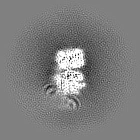
Journal: Sci Adv / Year: 2022 Title: Structural insight into UV-B-activated UVR8 bound to COP1. Authors: Yidong Wang / Lixia Wang / Zeyuan Guan / Hongfei Chang / Ling Ma / Cuicui Shen / Liang Qiu / Junjie Yan / Delin Zhang / Jian Li / Xing Wang Deng / Ping Yin / Abstract: The CONSTITUTIVE PHOTOMORPHOGENIC 1-SUPPRESSOR OF PHYA-105 (COP1-SPA) complex is a central repressor of photomorphogenesis. This complex acts as an E3 ubiquitin ligase downstream of various light ...The CONSTITUTIVE PHOTOMORPHOGENIC 1-SUPPRESSOR OF PHYA-105 (COP1-SPA) complex is a central repressor of photomorphogenesis. This complex acts as an E3 ubiquitin ligase downstream of various light signaling transduced from multiple photoreceptors in plants. How the COP1-SPA activity is regulated by divergent light-signaling pathways remains largely elusive. Here, we reproduced the regulation pathway of COP1-SPA in ultraviolet-B (UV-B) signaling in vitro and determined the cryo-electron microscopy structure of UV-B receptor UVR8 in complex with COP1. The complex formation is mediated by two-interface interactions between UV-B-activated UVR8 and COP1. Both interfaces are essential for the competitive binding of UVR8 against the signaling hub component HY5 to the COP1-SPA complex. We also show that RUP2 dissociates UVR8 from the COP1-SPA4-UVR8 complex and facilitates its redimerization. Our results support a UV-B signaling model that the COP1-SPA activity is repressed by UV-B-activated UVR8 and derepressed by RUP2, owing to competitive binding, and provide a framework for studying the regulatory roles of distinct photoreceptors on photomorphogenesis.
In the structure databanks used in Yorodumi, some data are registered as the other names, "COVID-19 virus" and "2019-nCoV". Here are the details of the virus and the list of structure data.
Jan 31, 2019. EMDB accession codes are about to change! (news from PDBe EMDB page)
EMDB accession codes are about to change! (news from PDBe EMDB page)
The allocation of 4 digits for EMDB accession codes will soon come to an end. Whilst these codes will remain in use, new EMDB accession codes will include an additional digit and will expand incrementally as the available range of codes is exhausted. The current 4-digit format prefixed with “EMD-” (i.e. EMD-XXXX) will advance to a 5-digit format (i.e. EMD-XXXXX), and so on. It is currently estimated that the 4-digit codes will be depleted around Spring 2019, at which point the 5-digit format will come into force.
The EM Navigator/Yorodumi systems omit the EMD- prefix.
Related info.:Q: What is EMD? / ID/Accession-code notation in Yorodumi/EM Navigator
Yorodumi is a browser for structure data from EMDB, PDB, SASBDB, etc.
This page is also the successor to EM Navigator detail page, and also detail information page/front-end page for Omokage search.
The word "yorodu" (or yorozu) is an old Japanese word meaning "ten thousand". "mi" (miru) is to see.
Related info.:EMDB / PDB / SASBDB / Comparison of 3 databanks / Yorodumi Search / Aug 31, 2016. New EM Navigator & Yorodumi / Yorodumi Papers / Jmol/JSmol / Function and homology information / Changes in new EM Navigator and Yorodumi
 Movie
Movie Controller

 Yorodumi
 Open data
 Basic information
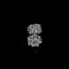
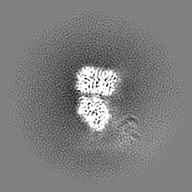
 Map data
 Sample
 Keywords
 Function and homology information
 Authors
 China, 1 items
  Citation
 Structure visualization
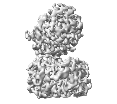
 Downloads & links
 emd_31968.png
 http://ftp.pdbj.org/pub/emdb/structures/EMD-31968
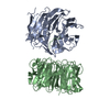


 Z (Sec.)
 Y (Row.)
 X (Col.)


 Sample components
 Homo sapiens (human)
 Processing
 Electron microscopy
 FIELD EMISSION GUN
