 Movie
Movie Controller

[English] 日本語
 Yorodumi
- PDB-7vef: The structure of GfsA KSQ-AT didomain in complex with a malonate ... -
+ Open data

- Basic information
| Entry | Database: PDB / ID: 7vef | ||||||
|---|---|---|---|---|---|---|---|
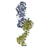
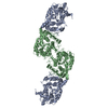
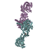
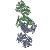
| Title | The structure of GfsA KSQ-AT didomain in complex with a malonate substrate analog | ||||||
 Components Components | Polyketide synthase | ||||||
 Keywords Keywords | TRANSFERASE / Decarboxylase / Thiolase fold / Polyketide biosynthesis / BIOSYNTHETIC PROTEIN | ||||||
| Function / homology | N-(2-acetamidoethyl)-2-nitro-ethanamide Function and homology information Function and homology information | ||||||
| Biological species |  Streptomyces graminofaciens (bacteria) Streptomyces graminofaciens (bacteria) | ||||||
| Method |  X-RAY DIFFRACTION / SYNCHROTRON / MOLECULAR REPLACEMENT / Resolution: 2.65 Å X-RAY DIFFRACTION / SYNCHROTRON / MOLECULAR REPLACEMENT / Resolution: 2.65 Å | ||||||
 Authors Authors | Chisuga, T. / Miyanaga, A. / Nagai, A. / Kudo, F. / Eguchi, T. | ||||||
| Funding support |  Japan, 1items Japan, 1items
| ||||||
 Citation Citation | Journal: Acs Chem.Biol. / Year: 2022 Title: Structural Insight into the Reaction Mechanism of Ketosynthase-Like Decarboxylase in a Loading Module of Modular Polyketide Synthases. Authors: Chisuga, T. / Nagai, A. / Miyanaga, A. / Goto, E. / Kishikawa, K. / Kudo, F. / Eguchi, T. | ||||||
| History |
|
- Structure visualization
| Structure viewer | Molecule: MolmilJmol/JSmol |
|---|
- Downloads & links
-Download
| PDBx/mmCIF format | 7vef.cif.gz | 312.8 KB | Display | PDBx/mmCIF format |
|---|---|---|---|---|
| PDB format | pdb7vef.ent.gz | 252 KB | Display | PDB format |
| PDBx/mmJSON format | 7vef.json.gz | Tree view | PDBx/mmJSON format | |
| Others |  Other downloads Other downloads |
-Validation report
| Arichive directory | https://data.pdbj.org/pub/pdb/validation_reports/ve/7vefftp://data.pdbj.org/pub/pdb/validation_reports/ve/7vef | HTTPS FTP |
|---|
-Related structure data
| Related structure data |  7veeSC S: Starting model for refinement C: citing same article ( |
|---|---|
| Similar structure data |


-Links
 PDBj
PDBj- Assembly
| Deposited unit | 
| |||||||||
|---|---|---|---|---|---|---|---|---|---|---|
| 1 | 
| |||||||||
| Unit cell |
| |||||||||
| Components on special symmetry positions |
|
-Components
| #1: Protein | Mass: 95532.602 Da / Num. of mol.: 1 Source method: isolated from a genetically manipulated source Source: (gene. exp.) Streptomyces graminofaciens (bacteria) / Gene: gfsA / Plasmid: pCold III / Production host: |
|---|---|
| #2: Chemical | ChemComp-6IU /   Mass: 189.169 Da / Num. of mol.: 1 / Source method: obtained synthetically / Formula: C6H11N3O4 / Feature type: SUBJECT OF INVESTIGATION Mass: 189.169 Da / Num. of mol.: 1 / Source method: obtained synthetically / Formula: C6H11N3O4 / Feature type: SUBJECT OF INVESTIGATION |
| #3: Chemical | ChemComp-GOL /   Mass: 92.094 Da / Num. of mol.: 1 / Source method: obtained synthetically / Formula: C3H8O3 Mass: 92.094 Da / Num. of mol.: 1 / Source method: obtained synthetically / Formula: C3H8O3 |
| #4: Water | ChemComp-HOH /  Mass: 18.015 Da / Num. of mol.: 27 / Source method: isolated from a natural source / Formula: H2O Mass: 18.015 Da / Num. of mol.: 27 / Source method: isolated from a natural source / Formula: H2O |
| Has ligand of interest | Y |
-Experimental details
-Experiment
| Experiment | Method: X-RAY DIFFRACTION / Number of used crystals: 1 |
|---|
- Sample preparation
| Crystal | Density Matthews: 3.17 Å3/Da / Density % sol: 61.22 % |
|---|---|
| Crystal grow | Temperature: 298 K / Method: vapor diffusion, sitting drop / pH: 6.1 / Details: sodium malonate, glycerol, sodium chloride, HEPES |
-Data collection
| Diffraction | Mean temperature: 100 K / Serial crystal experiment: N |
|---|---|
| Diffraction source | Source: SYNCHROTRON / Site: Photon Factory / Beamline: BL-5A / Wavelength: 1 Å |
| Detector | Type: DECTRIS PILATUS3 S 6M / Detector: PIXEL / Date: May 29, 2021 |
| Radiation | Monochromator: Numerical link type Si(111) double crystal monochromator Protocol: SINGLE WAVELENGTH / Monochromatic (M) / Laue (L): M / Scattering type: x-ray |
| Radiation wavelength | Wavelength: 1 Å / Relative weight: 1 |
| Reflection | Resolution: 2.65→50 Å / Num. obs: 34710 / % possible obs: 99.3 % / Redundancy: 6.3 % / CC1/2: 0.998 / Rmerge(I) obs: 0.063 / Net I/σ(I): 16.1 |
| Reflection shell | Resolution: 2.65→2.79 Å / Redundancy: 6.4 % / Rmerge(I) obs: 0.701 / Mean I/σ(I) obs: 2.4 / Num. unique obs: 5006 / CC1/2: 0.857 / % possible all: 99.1 |
- Processing
| Software |
| ||||||||||||||||||||||||||||||||||||||||||||||||||||||||||||
|---|---|---|---|---|---|---|---|---|---|---|---|---|---|---|---|---|---|---|---|---|---|---|---|---|---|---|---|---|---|---|---|---|---|---|---|---|---|---|---|---|---|---|---|---|---|---|---|---|---|---|---|---|---|---|---|---|---|---|---|---|---|
| Refinement | Method to determine structure: MOLECULAR REPLACEMENT Starting model: 7VEE Resolution: 2.65→46.63 Å / Cor.coef. Fo:Fc: 0.957 / Cor.coef. Fo:Fc free: 0.925 / SU B: 31.087 / SU ML: 0.274 / Cross valid method: THROUGHOUT / σ(F): 0 / ESU R: 0.464 / ESU R Free: 0.298 / Stereochemistry target values: MAXIMUM LIKELIHOOD Details: U VALUES : WITH TLS ADDED HYDROGENS HAVE BEEN ADDED IN THE RIDING POSITIONS
| ||||||||||||||||||||||||||||||||||||||||||||||||||||||||||||
| Solvent computation | Ion probe radii: 0.8 Å / Shrinkage radii: 0.8 Å / VDW probe radii: 1.2 Å / Solvent model: MASK | ||||||||||||||||||||||||||||||||||||||||||||||||||||||||||||
| Displacement parameters | Biso max: 133.68 Å2 / Biso mean: 75.738 Å2 / Biso min: 39.52 Å2
| ||||||||||||||||||||||||||||||||||||||||||||||||||||||||||||
| Refinement step | Cycle: final / Resolution: 2.65→46.63 Å
| ||||||||||||||||||||||||||||||||||||||||||||||||||||||||||||
| Refine LS restraints |
| ||||||||||||||||||||||||||||||||||||||||||||||||||||||||||||
| LS refinement shell | Resolution: 2.65→2.719 Å / Rfactor Rfree error: 0 / Total num. of bins used: 20
| ||||||||||||||||||||||||||||||||||||||||||||||||||||||||||||
| Refinement TLS params. | Method: refined / Origin x: -61.745 Å / Origin y: 33.905 Å / Origin z: 1.196 Å
| ||||||||||||||||||||||||||||||||||||||||||||||||||||||||||||
| Refinement TLS group |
|
