 Movie
Movie Controller

[English] 日本語
 Yorodumi
- PDB-7vc7: The structure of beta-xylosidase from Phanerochaete chrysosporium... -
+ Open data

- Basic information
| Entry | Database: PDB / ID: 7vc7 | ||||||||||||
|---|---|---|---|---|---|---|---|---|---|---|---|---|---|
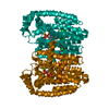
| Title | The structure of beta-xylosidase from Phanerochaete chrysosporium(PcBxl3) | ||||||||||||
 Components Components | xylan 1,4-beta-xylosidase | ||||||||||||
 Keywords Keywords | HYDROLASE / Glycoside hydrolase family 3 / beta-xylosidase / HYDROLASE (E.C.3.2.1.37) | ||||||||||||
| Function / homology |  Function and homology information Function and homology informationGlycoside hydrolase family 3 C-terminal domain / Glycoside hydrolase, family 3, N-terminal domain / TIM Barrel / Alpha-Beta Barrel / Immunoglobulins / Immunoglobulin-like / Sandwich / Rossmann fold / 3-Layer(aba) Sandwich / Mainly Beta / Alpha Beta Similarity search - Domain/homology | ||||||||||||
| Biological species |  Phanerochaete chrysosporium (fungus) Phanerochaete chrysosporium (fungus) | ||||||||||||
| Method |  X-RAY DIFFRACTION / SYNCHROTRON / MOLECULAR REPLACEMENT / Resolution: 3.08 Å X-RAY DIFFRACTION / SYNCHROTRON / MOLECULAR REPLACEMENT / Resolution: 3.08 Å | ||||||||||||
 Authors Authors | Kojima, K. / Sunagawa, N. / Igarashi, K. | ||||||||||||
| Funding support |  Japan, 3items Japan, 3items
| ||||||||||||
 Citation Citation | Journal: J.Biol.Chem. / Year: 2022 Title: Comparison of glycoside hydrolase family 3 beta-xylosidases from basidiomycetes and ascomycetes reveals evolutionarily distinct xylan degradation systems. Authors: Kojima, K. / Sunagawa, N. / Mikkelsen, N.E. / Hansson, H. / Karkehabadi, S. / Samejima, M. / Sandgren, M. / Igarashi, K. | ||||||||||||
| History |
|
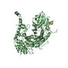
- Structure visualization
| Structure viewer | Molecule: MolmilJmol/JSmol |
|---|
- Downloads & links
-Download
| PDBx/mmCIF format | 7vc7.cif.gz | 166 KB | Display | PDBx/mmCIF format |
|---|---|---|---|---|
| PDB format | pdb7vc7.ent.gz | 128.6 KB | Display | PDB format |
| PDBx/mmJSON format | 7vc7.json.gz | Tree view | PDBx/mmJSON format | |
| Others |  Other downloads Other downloads |
-Validation report
| Arichive directory | https://data.pdbj.org/pub/pdb/validation_reports/vc/7vc7ftp://data.pdbj.org/pub/pdb/validation_reports/vc/7vc7 | HTTPS FTP |
|---|
-Related structure data
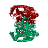
| Related structure data | 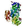 7vc6SC S: Starting model for refinement C: citing same article ( |
|---|---|
| Similar structure data |


-Links
 PDBj
PDBj- Assembly
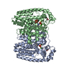
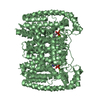
| Deposited unit | 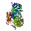
| ||||||||||
|---|---|---|---|---|---|---|---|---|---|---|---|
| 1 |
| ||||||||||
| Unit cell |
|
-Components
-Protein , 1 types, 1 molecules A
| #1: Protein | Mass: 79513.883 Da / Num. of mol.: 1 Source method: isolated from a genetically manipulated source Source: (gene. exp.) Phanerochaete chrysosporium (fungus) / Strain: K-3 / Gene: PcBxl3 / Plasmid: pPICZalphaA / Production host: Komagataella pastoris (fungus) / Strain (production host): KM71H / References: xylan 1,4-beta-xylosidase |
|---|
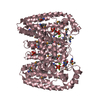
-Sugars , 2 types, 8 molecules 
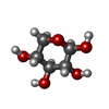
| #5: Sugar | ChemComp-NAG /  Type: D-saccharide, beta linking / Mass: 221.208 Da / Num. of mol.: 7 Type: D-saccharide, beta linking / Mass: 221.208 Da / Num. of mol.: 7Source method: isolated from a genetically manipulated source Formula: C8H15NO6 #6: Sugar | ChemComp-XYP / |  Type: D-saccharide, beta linking / Mass: 150.130 Da / Num. of mol.: 1 Type: D-saccharide, beta linking / Mass: 150.130 Da / Num. of mol.: 1Source method: isolated from a genetically manipulated source Formula: C5H10O5 / Feature type: SUBJECT OF INVESTIGATION |
|---|
-Non-polymers , 4 types, 186 molecules 


| #2: Chemical | ChemComp-1PE /  Mass: 238.278 Da / Num. of mol.: 12 / Source method: obtained synthetically / Formula: C10H22O6 / Comment: precipitant*YM Mass: 238.278 Da / Num. of mol.: 12 / Source method: obtained synthetically / Formula: C10H22O6 / Comment: precipitant*YM#3: Chemical | ChemComp-PEG /  Mass: 106.120 Da / Num. of mol.: 5 / Source method: obtained synthetically / Formula: C4H10O3 Mass: 106.120 Da / Num. of mol.: 5 / Source method: obtained synthetically / Formula: C4H10O3#4: Chemical | ChemComp-EDO / |  Mass: 62.068 Da / Num. of mol.: 1 / Source method: obtained synthetically / Formula: C2H6O2 Mass: 62.068 Da / Num. of mol.: 1 / Source method: obtained synthetically / Formula: C2H6O2#7: Water | ChemComp-HOH / | Mass: 18.015 Da / Num. of mol.: 168 / Source method: isolated from a natural source / Formula: H2O |
|---|
-Details
| Has ligand of interest | Y |
|---|---|
| Has protein modification | Y |
-Experimental details
-Experiment
| Experiment | Method: X-RAY DIFFRACTION / Number of used crystals: 1 |
|---|
- Sample preparation
| Crystal | Density Matthews: 2.41 Å3/Da / Density % sol: 48.95 % |
|---|---|
| Crystal grow | Temperature: 293 K / Method: vapor diffusion, sitting drop / pH: 7 / Details: 525mM Malic acid (pH7.0), 20% v/v PEG 3350 |
-Data collection
| Diffraction | Mean temperature: 95 K / Serial crystal experiment: N |
|---|---|
| Diffraction source | Source: SYNCHROTRON / Site: Photon Factory / Beamline: BL-5A / Wavelength: 1 Å |
| Detector | Type: DECTRIS PILATUS3 S 6M / Detector: PIXEL / Date: Dec 22, 2020 |
| Radiation | Protocol: SINGLE WAVELENGTH / Monochromatic (M) / Laue (L): M / Scattering type: x-ray |
| Radiation wavelength | Wavelength: 1 Å / Relative weight: 1 |
| Reflection | Resolution: 3.08→44.08 Å / Num. obs: 14756 / % possible obs: 99.91 % / Redundancy: 2 % / Biso Wilson estimate: 48.53 Å2 / CC1/2: 0.985 / Net I/σ(I): 7.82 |
| Reflection shell | Resolution: 3.08→3.19 Å / Num. unique obs: 1450 / CC1/2: 0.8 |
- Processing
| Software |
| ||||||||||||||||||||||||||||||||||||||||||
|---|---|---|---|---|---|---|---|---|---|---|---|---|---|---|---|---|---|---|---|---|---|---|---|---|---|---|---|---|---|---|---|---|---|---|---|---|---|---|---|---|---|---|---|
| Refinement | Method to determine structure: MOLECULAR REPLACEMENT Starting model: 7VC6 Resolution: 3.08→44.079 Å / SU ML: 0.34 / Cross valid method: FREE R-VALUE / σ(F): 1.34 / Phase error: 25.31 / Stereochemistry target values: ML
| ||||||||||||||||||||||||||||||||||||||||||
| Solvent computation | Shrinkage radii: 0.9 Å / VDW probe radii: 1.11 Å / Solvent model: FLAT BULK SOLVENT MODEL | ||||||||||||||||||||||||||||||||||||||||||
| Refinement step | Cycle: LAST / Resolution: 3.08→44.079 Å
| ||||||||||||||||||||||||||||||||||||||||||
| Refine LS restraints |
| ||||||||||||||||||||||||||||||||||||||||||
| LS refinement shell |
|
