 Movie
Movie Controller

[English] 日本語
 Yorodumi
- PDB-4rxd: T. Brucei Farnesyl Diphosphate Synthase Complexed with Risedronate -
+ Open data

- Basic information
| Entry | Database: PDB / ID: 4rxd | ||||||
|---|---|---|---|---|---|---|---|
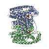
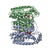
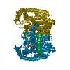
| Title | T. Brucei Farnesyl Diphosphate Synthase Complexed with Risedronate | ||||||
 Components Components | Farnesyl pyrophosphate synthase | ||||||
 Keywords Keywords | TRANSFERASE | ||||||
| Function / homology |  Function and homology information Function and homology informationtrans, trans-farnesyl diphosphate biosynthetic process / dimethylallyltranstransferase activity / (2E,6E)-farnesyl diphosphate synthase activity / metal ion binding / cytoplasm Similarity search - Function | ||||||
| Biological species |  | ||||||
| Method |  X-RAY DIFFRACTION / SYNCHROTRON / MOLECULAR REPLACEMENT / Resolution: 2 Å X-RAY DIFFRACTION / SYNCHROTRON / MOLECULAR REPLACEMENT / Resolution: 2 Å | ||||||
 Authors Authors | Cao, R. / Liu, Y.-L. / Oldfield, E. | ||||||
 Citation Citation | Journal: ACS Med Chem Lett / Year: 2015 Title: Farnesyl diphosphate synthase inhibitors with unique ligand-binding geometries. Authors: Liu, Y.L. / Cao, R. / Wang, Y. / Oldfield, E. | ||||||
| History |
|
- Structure visualization
| Structure viewer | Molecule: MolmilJmol/JSmol |
|---|
- Downloads & links
-Download
| PDBx/mmCIF format | 4rxd.cif.gz | 234.3 KB | Display | PDBx/mmCIF format |
|---|---|---|---|---|
| PDB format | pdb4rxd.ent.gz | 186.6 KB | Display | PDB format |
| PDBx/mmJSON format | 4rxd.json.gz | Tree view | PDBx/mmJSON format | |
| Others |  Other downloads Other downloads |
-Validation report
| Arichive directory | https://data.pdbj.org/pub/pdb/validation_reports/rx/4rxdftp://data.pdbj.org/pub/pdb/validation_reports/rx/4rxd | HTTPS FTP |
|---|
-Related structure data
| Related structure data | 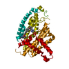 4rxaC  4rxcC  4rxeC  4rypC 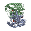 2ogdS C: citing same article ( S: Starting model for refinement |
|---|---|
| Similar structure data |


-Links
 PDBj
PDBj
- Assembly
| Deposited unit | 
| ||||||||
|---|---|---|---|---|---|---|---|---|---|
| 1 | 
| ||||||||
| 2 | 
| ||||||||
| Unit cell |
| ||||||||
| Components on special symmetry positions |
|
-Components
| #1: Protein | Mass: 44475.738 Da / Num. of mol.: 3 Source method: isolated from a genetically manipulated source Source: (gene. exp.)  #2: Chemical | ChemComp-MG /   Mass: 24.305 Da / Num. of mol.: 9 / Source method: obtained synthetically / Formula: Mg Mass: 24.305 Da / Num. of mol.: 9 / Source method: obtained synthetically / Formula: Mg#3: Chemical |   Mass: 283.112 Da / Num. of mol.: 3 / Source method: obtained synthetically / Formula: C7H11NO7P2 Mass: 283.112 Da / Num. of mol.: 3 / Source method: obtained synthetically / Formula: C7H11NO7P2#4: Water | ChemComp-HOH / |  Mass: 18.015 Da / Num. of mol.: 484 / Source method: isolated from a natural source / Formula: H2O Mass: 18.015 Da / Num. of mol.: 484 / Source method: isolated from a natural source / Formula: H2O |
|---|
-Experimental details
-Experiment
| Experiment | Method: X-RAY DIFFRACTION / Number of used crystals: 1 |
|---|
- Sample preparation
| Crystal | Density Matthews: 2.35 Å3/Da / Density % sol: 47.58 % |
|---|---|
| Crystal grow | Temperature: 298 K / Method: vapor diffusion, hanging drop / pH: 5.75 Details: 10% MPD, 0.1 AMMONIUM ACETATE, pH 5.75, VAPOR DIFFUSION, HANGING DROP, temperature 298K |
-Data collection
| Diffraction | Mean temperature: 298 K |
|---|---|
| Diffraction source | Source: SYNCHROTRON / Site: APS  / Beamline: 21-ID-G / Wavelength: 0.97857 Å / Beamline: 21-ID-G / Wavelength: 0.97857 Å |
| Detector | Type: MARMOSAIC 300 mm CCD / Detector: CCD / Date: Apr 28, 2008 |
| Radiation | Monochromator: C(111) / Protocol: SINGLE WAVELENGTH / Monochromatic (M) / Laue (L): M / Scattering type: x-ray |
| Radiation wavelength | Wavelength: 0.97857 Å / Relative weight: 1 |
| Reflection | Resolution: 2→50 Å / Num. all: 82972 / % possible obs: 99.8 % / Observed criterion σ(F): 1 / Observed criterion σ(I): 1 |
| Reflection shell | Resolution: 2→2.07 Å / % possible all: 99.2 |
- Processing
| Software |
| |||||||||||||||||||||||||||||||||||||||||||||||||||||||||||||||||||||||||||||||||||||||||||||||
|---|---|---|---|---|---|---|---|---|---|---|---|---|---|---|---|---|---|---|---|---|---|---|---|---|---|---|---|---|---|---|---|---|---|---|---|---|---|---|---|---|---|---|---|---|---|---|---|---|---|---|---|---|---|---|---|---|---|---|---|---|---|---|---|---|---|---|---|---|---|---|---|---|---|---|---|---|---|---|---|---|---|---|---|---|---|---|---|---|---|---|---|---|---|---|---|---|
| Refinement | Method to determine structure: MOLECULAR REPLACEMENT Starting model: PDB ENTRY 2OGD Resolution: 2→29.55 Å / Cor.coef. Fo:Fc: 0.941 / Cor.coef. Fo:Fc free: 0.919 / SU B: 4.137 / SU ML: 0.119 / Cross valid method: THROUGHOUT / σ(F): 0 / ESU R: 0.2 / ESU R Free: 0.174 / Stereochemistry target values: MAXIMUM LIKELIHOOD / Details: HYDROGENS HAVE BEEN ADDED IN THE RIDING POSITIONS
| |||||||||||||||||||||||||||||||||||||||||||||||||||||||||||||||||||||||||||||||||||||||||||||||
| Solvent computation | Ion probe radii: 0.8 Å / Shrinkage radii: 0.8 Å / VDW probe radii: 1.4 Å / Solvent model: BABINET MODEL WITH MASK | |||||||||||||||||||||||||||||||||||||||||||||||||||||||||||||||||||||||||||||||||||||||||||||||
| Displacement parameters | Biso max: 84.9 Å2 / Biso mean: 28.582 Å2 / Biso min: 10.13 Å2
| |||||||||||||||||||||||||||||||||||||||||||||||||||||||||||||||||||||||||||||||||||||||||||||||
| Refinement step | Cycle: LAST / Resolution: 2→29.55 Å
| |||||||||||||||||||||||||||||||||||||||||||||||||||||||||||||||||||||||||||||||||||||||||||||||
| Refine LS restraints |
| |||||||||||||||||||||||||||||||||||||||||||||||||||||||||||||||||||||||||||||||||||||||||||||||
| LS refinement shell | Resolution: 2.001→2.053 Å / Total num. of bins used: 20
|
