 Movie
Movie Controller

[English] 日本語
 Yorodumi
- PDB-7v8t: Crystal structure of class II pyruvate aldolase from Pseudomonas ... -
+ Open data

- Basic information
| Entry | Database: PDB / ID: 7v8t | ||||||
|---|---|---|---|---|---|---|---|
| Title | Crystal structure of class II pyruvate aldolase from Pseudomonas aeruginosa. | ||||||
 Components Components | 2,4-dihydroxyhept-2-ene-1,7-dioic acid aldolase | ||||||
 Keywords Keywords | LYASE / Class II pyruvate aldolase / Hexamer / TIM barrel fold | ||||||
| Function / homology |  Function and homology information Function and homology information: / Lyases; Carbon-carbon lyases; Aldehyde-lyases / phenylacetate catabolic process / metal ion binding Similarity search - Function | ||||||
| Biological species |   Pseudomonas aeruginosa (bacteria) Pseudomonas aeruginosa (bacteria) | ||||||
| Method |  X-RAY DIFFRACTION / SYNCHROTRON / MOLECULAR REPLACEMENT / Resolution: 2.48 Å X-RAY DIFFRACTION / SYNCHROTRON / MOLECULAR REPLACEMENT / Resolution: 2.48 Å | ||||||
 Authors Authors | Seo, P.W. / Kim, J.S. | ||||||
| Funding support |  Korea, Republic Of, 1items Korea, Republic Of, 1items
| ||||||
 Citation Citation | Journal: To Be Published Title: Biochemical and Molecular Characterization of Pyruvate Aldolase for the Synthesis of 2-Keto-4-hydroxybutyrate. Authors: Jeong, Y.J. / Le, T.K. / Seo, P.W. / Ju, S.B. / Kim, J.S. / Yeom, S.J. | ||||||
| History |
|
- Structure visualization
| Structure viewer | Molecule: MolmilJmol/JSmol |
|---|
- Downloads & links
-Download
| PDBx/mmCIF format | 7v8t.cif.gz | 565.4 KB | Display | PDBx/mmCIF format |
|---|---|---|---|---|
| PDB format | pdb7v8t.ent.gz | 466.3 KB | Display | PDB format |
| PDBx/mmJSON format | 7v8t.json.gz | Tree view | PDBx/mmJSON format | |
| Others |  Other downloads Other downloads |
-Validation report
| Arichive directory | https://data.pdbj.org/pub/pdb/validation_reports/v8/7v8tftp://data.pdbj.org/pub/pdb/validation_reports/v8/7v8t | HTTPS FTP |
|---|
-Related structure data
| Related structure data | 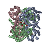 2vwtS S: Starting model for refinement |
|---|---|
| Similar structure data |
-Links
 PDBj
PDBj
- Assembly
| Deposited unit | 
| |||||||||||||||||||||||||||||||||||||||||||||||||||||||||||||||
|---|---|---|---|---|---|---|---|---|---|---|---|---|---|---|---|---|---|---|---|---|---|---|---|---|---|---|---|---|---|---|---|---|---|---|---|---|---|---|---|---|---|---|---|---|---|---|---|---|---|---|---|---|---|---|---|---|---|---|---|---|---|---|---|---|
| 1 |
| |||||||||||||||||||||||||||||||||||||||||||||||||||||||||||||||
| Unit cell |
| |||||||||||||||||||||||||||||||||||||||||||||||||||||||||||||||
| Noncrystallographic symmetry (NCS) | NCS domain:
NCS domain segments:
|
-Components
| #1: Protein | Mass: 28230.148 Da / Num. of mol.: 6 Source method: isolated from a genetically manipulated source Source: (gene. exp.) Pseudomonas aeruginosa (bacteria) / Production host: References: UniProt: A0A081HJP9, 4-hydroxy-2-oxoheptanedioate aldolase #2: Chemical | ChemComp-IOD /   Mass: 126.904 Da / Num. of mol.: 10 / Source method: obtained synthetically / Formula: I / Feature type: SUBJECT OF INVESTIGATION Mass: 126.904 Da / Num. of mol.: 10 / Source method: obtained synthetically / Formula: I / Feature type: SUBJECT OF INVESTIGATION#3: Chemical |   Mass: 282.331 Da / Num. of mol.: 2 / Source method: obtained synthetically / Formula: C12H26O7 / Feature type: SUBJECT OF INVESTIGATION / Comment: precipitant*YM Mass: 282.331 Da / Num. of mol.: 2 / Source method: obtained synthetically / Formula: C12H26O7 / Feature type: SUBJECT OF INVESTIGATION / Comment: precipitant*YM#4: Water | ChemComp-HOH / |  Mass: 18.015 Da / Num. of mol.: 317 / Source method: isolated from a natural source / Formula: H2O Mass: 18.015 Da / Num. of mol.: 317 / Source method: isolated from a natural source / Formula: H2OHas ligand of interest | Y | Has protein modification | Y | |
|---|
-Experimental details
-Experiment
| Experiment | Method: X-RAY DIFFRACTION / Number of used crystals: 1 |
|---|
- Sample preparation
| Crystal | Density Matthews: 2.28 Å3/Da / Density % sol: 45.94 % |
|---|---|
| Crystal grow | Temperature: 294 K / Method: vapor diffusion, hanging drop / Details: 200mM sodium iodide, 24% (w/v) PEG 3350 |
-Data collection
| Diffraction | Mean temperature: 100 K / Serial crystal experiment: N |
|---|---|
| Diffraction source | Source: SYNCHROTRON / Site: PAL/PLS / Beamline: 11C / Wavelength: 0.9794 Å |
| Detector | Type: DECTRIS PILATUS 6M / Detector: PIXEL / Date: Mar 12, 2020 |
| Radiation | Protocol: SINGLE WAVELENGTH / Monochromatic (M) / Laue (L): M / Scattering type: x-ray |
| Radiation wavelength | Wavelength: 0.9794 Å / Relative weight: 1 |
| Reflection | Resolution: 2.48→50 Å / Num. obs: 51389 / % possible obs: 96.6 % / Redundancy: 6 % / Biso Wilson estimate: 39.26 Å2 / Rpim(I) all: 0.094 / Rrim(I) all: 0.241 / Rsym value: 0.173 / Net I/σ(I): 6.4 |
| Reflection shell | Resolution: 2.5→2.54 Å / Num. unique obs: 2440 / CC1/2: 0.468 / Rpim(I) all: 0.566 / Rsym value: 0.856 |
- Processing
| Software |
| ||||||||||||||||||||||||||||||||||||||||||||||||||||||||||||||||||||||||||||||||||||||||||||||||||||||||||||||||
|---|---|---|---|---|---|---|---|---|---|---|---|---|---|---|---|---|---|---|---|---|---|---|---|---|---|---|---|---|---|---|---|---|---|---|---|---|---|---|---|---|---|---|---|---|---|---|---|---|---|---|---|---|---|---|---|---|---|---|---|---|---|---|---|---|---|---|---|---|---|---|---|---|---|---|---|---|---|---|---|---|---|---|---|---|---|---|---|---|---|---|---|---|---|---|---|---|---|---|---|---|---|---|---|---|---|---|---|---|---|---|---|---|---|
| Refinement | Method to determine structure: MOLECULAR REPLACEMENT Starting model: 2VWT Resolution: 2.48→46.54 Å / SU ML: 0.31 / Cross valid method: THROUGHOUT / σ(F): 1.35 / Phase error: 22.37 / Stereochemistry target values: ML
| ||||||||||||||||||||||||||||||||||||||||||||||||||||||||||||||||||||||||||||||||||||||||||||||||||||||||||||||||
| Solvent computation | Shrinkage radii: 0.9 Å / VDW probe radii: 1.11 Å / Solvent model: FLAT BULK SOLVENT MODEL | ||||||||||||||||||||||||||||||||||||||||||||||||||||||||||||||||||||||||||||||||||||||||||||||||||||||||||||||||
| Displacement parameters | Biso max: 115.11 Å2 / Biso mean: 42.2375 Å2 / Biso min: 20.45 Å2 | ||||||||||||||||||||||||||||||||||||||||||||||||||||||||||||||||||||||||||||||||||||||||||||||||||||||||||||||||
| Refinement step | Cycle: final / Resolution: 2.48→46.54 Å
| ||||||||||||||||||||||||||||||||||||||||||||||||||||||||||||||||||||||||||||||||||||||||||||||||||||||||||||||||
| Refine LS restraints NCS |
| ||||||||||||||||||||||||||||||||||||||||||||||||||||||||||||||||||||||||||||||||||||||||||||||||||||||||||||||||
| LS refinement shell | Refine-ID: X-RAY DIFFRACTION / Rfactor Rfree error: 0 / Total num. of bins used: 15
| ||||||||||||||||||||||||||||||||||||||||||||||||||||||||||||||||||||||||||||||||||||||||||||||||||||||||||||||||
| Refinement TLS params. | Method: refined / Origin x: -19.1135 Å / Origin y: -19.6095 Å / Origin z: 4.9468 Å
| ||||||||||||||||||||||||||||||||||||||||||||||||||||||||||||||||||||||||||||||||||||||||||||||||||||||||||||||||
| Refinement TLS group |
|
