 Movie
Movie Controller

[English] 日本語
 Yorodumi
- PDB-7v8e: Crystal structure of IpaH1.4 LRR domain bound to HOIL-1L UBL domain. -
+ Open data

- Basic information
| Entry | Database: PDB / ID: 7v8e | ||||||||||||||||||
|---|---|---|---|---|---|---|---|---|---|---|---|---|---|---|---|---|---|---|---|
| Title | Crystal structure of IpaH1.4 LRR domain bound to HOIL-1L UBL domain. | ||||||||||||||||||
 Components Components |
| ||||||||||||||||||
 Keywords Keywords | LIGASE / E3 liagse / ubiquitin / innate immune | ||||||||||||||||||
| Function / homology |  Function and homology information Function and homology informationmodulation of process of another organism / protein linear polyubiquitination / LUBAC complex / RBR-type E3 ubiquitin transferase / negative regulation of necroptotic process / ubiquitin ligase activator activity / : / TNFR1-induced proapoptotic signaling / positive regulation of extrinsic apoptotic signaling pathway / canonical NF-kappaB signal transduction ...modulation of process of another organism / protein linear polyubiquitination / LUBAC complex / RBR-type E3 ubiquitin transferase / negative regulation of necroptotic process / ubiquitin ligase activator activity / : / TNFR1-induced proapoptotic signaling / positive regulation of extrinsic apoptotic signaling pathway / canonical NF-kappaB signal transduction / : / negative regulation of canonical NF-kappaB signal transduction / TNFR1-induced NF-kappa-B signaling pathway / ubiquitin binding / protein sequestering activity / Regulation of TNFR1 signaling / positive regulation of non-canonical NF-kappaB signal transduction / RING-type E3 ubiquitin transferase / protein polyubiquitination / ubiquitin-protein transferase activity / ubiquitin protein ligase activity / Antigen processing: Ubiquitination & Proteasome degradation / T cell receptor signaling pathway / host cell cytoplasm / proteasome-mediated ubiquitin-dependent protein catabolic process / positive regulation of canonical NF-kappaB signal transduction / transcription coactivator activity / defense response to bacterium / protein ubiquitination / extracellular region / zinc ion binding / identical protein binding / cytosol Similarity search - Function | ||||||||||||||||||
| Biological species |  Shigella flexneri 5a str. M90T (bacteria) Shigella flexneri 5a str. M90T (bacteria) Homo sapiens (human) Homo sapiens (human) | ||||||||||||||||||
| Method |  X-RAY DIFFRACTION / SYNCHROTRON / MOLECULAR REPLACEMENT / Resolution: 1.9 Å X-RAY DIFFRACTION / SYNCHROTRON / MOLECULAR REPLACEMENT / Resolution: 1.9 Å | ||||||||||||||||||
 Authors Authors | Liu, J. / Wang, Y. / Pan, L. | ||||||||||||||||||
| Funding support |  China, 5items China, 5items
| ||||||||||||||||||
 Citation Citation | Journal: Proc.Natl.Acad.Sci.USA / Year: 2022 Title: Mechanistic insights into the subversion of the linear ubiquitin chain assembly complex by the E3 ligase IpaH1.4 of Shigella flexneri. Authors: Liu, J. / Wang, Y. / Wang, D. / Wang, Y. / Xu, X. / Zhang, Y. / Li, Y. / Zhang, M. / Gong, X. / Tang, Y. / Shen, L. / Li, M. / Pan, L. | ||||||||||||||||||
| History |
|
- Structure visualization
| Structure viewer | Molecule: MolmilJmol/JSmol |
|---|
- Downloads & links
-Download
| PDBx/mmCIF format | 7v8e.cif.gz | 176.2 KB | Display | PDBx/mmCIF format |
|---|---|---|---|---|
| PDB format | pdb7v8e.ent.gz | 110 KB | Display | PDB format |
| PDBx/mmJSON format | 7v8e.json.gz | Tree view | PDBx/mmJSON format | |
| Others |  Other downloads Other downloads |
-Validation report
| Arichive directory | https://data.pdbj.org/pub/pdb/validation_reports/v8/7v8eftp://data.pdbj.org/pub/pdb/validation_reports/v8/7v8e | HTTPS FTP |
|---|
-Related structure data
| Related structure data |  7v8fC 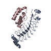 7v8gC 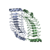 7v8hC  4dbgS S: Starting model for refinement C: citing same article ( |
|---|---|
| Similar structure data |
-Links
 PDBj
PDBj


- Assembly
| Deposited unit | 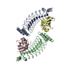
| ||||||||||||||||||||||||||||||||||||||||||||||||||||||||||||||||||||||||
|---|---|---|---|---|---|---|---|---|---|---|---|---|---|---|---|---|---|---|---|---|---|---|---|---|---|---|---|---|---|---|---|---|---|---|---|---|---|---|---|---|---|---|---|---|---|---|---|---|---|---|---|---|---|---|---|---|---|---|---|---|---|---|---|---|---|---|---|---|---|---|---|---|---|
| 1 | 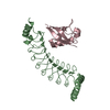
| ||||||||||||||||||||||||||||||||||||||||||||||||||||||||||||||||||||||||
| 2 | 
| ||||||||||||||||||||||||||||||||||||||||||||||||||||||||||||||||||||||||
| Unit cell |
| ||||||||||||||||||||||||||||||||||||||||||||||||||||||||||||||||||||||||
| Noncrystallographic symmetry (NCS) | NCS domain:
NCS domain segments:
NCS ensembles :
NCS oper:
|
-Components
| #1: Protein | Mass: 26991.988 Da / Num. of mol.: 2 Source method: isolated from a genetically manipulated source Source: (gene. exp.) Shigella flexneri 5a str. M90T (bacteria)Strain: M90T / Gene: ipaH1.4 / Production host: References: UniProt: Q9AFJ5, RING-type E3 ubiquitin transferase #2: Protein | Mass: 10183.492 Da / Num. of mol.: 2 Source method: isolated from a genetically manipulated source Source: (gene. exp.) Homo sapiens (human) / Production host: References: UniProt: Q9BYM8, RBR-type E3 ubiquitin transferase #3: Water | ChemComp-HOH / |  Mass: 18.015 Da / Num. of mol.: 431 / Source method: isolated from a natural source / Formula: H2O Mass: 18.015 Da / Num. of mol.: 431 / Source method: isolated from a natural source / Formula: H2O |
|---|
-Experimental details
-Experiment
| Experiment | Method: X-RAY DIFFRACTION / Number of used crystals: 1 |
|---|
- Sample preparation
| Crystal | Density Matthews: 2.12 Å3/Da / Density % sol: 42.09 % |
|---|---|
| Crystal grow | Temperature: 289 K / Method: evaporation / pH: 8.5 Details: 0.2 M (NH4)2SO4, 0.1 M Tris-HCl (pH 8.5), and 25% (w/v) PEG3350 |
-Data collection
| Diffraction | Mean temperature: 100 K / Serial crystal experiment: N |
|---|---|
| Diffraction source | Source: SYNCHROTRON / Site: SSRF / Beamline: BL17U1 / Wavelength: 0.9792 Å |
| Detector | Type: DECTRIS EIGER X 16M / Detector: PIXEL / Date: Dec 15, 2019 |
| Radiation | Protocol: SINGLE WAVELENGTH / Monochromatic (M) / Laue (L): M / Scattering type: x-ray |
| Radiation wavelength | Wavelength: 0.9792 Å / Relative weight: 1 |
| Reflection | Resolution: 1.9→49.38 Å / Num. obs: 47800 / % possible obs: 99.8 % / Redundancy: 6.3 % / Biso Wilson estimate: 34.14 Å2 / CC1/2: 0.998 / Rpim(I) all: 0.025 / Net I/σ(I): 16.3 |
| Reflection shell | Resolution: 1.9→1.94 Å / Mean I/σ(I) obs: 2.3 / Num. unique obs: 3094 / CC1/2: 0.765 / Rpim(I) all: 0.283 / % possible all: 99.5 |
- Processing
| Software |
| ||||||||||||||||||||||||||||||||||||||||||||||||||||||||||||||||||||||||||||||||||||||||||||||||||||||||||||||||||||||||||||||
|---|---|---|---|---|---|---|---|---|---|---|---|---|---|---|---|---|---|---|---|---|---|---|---|---|---|---|---|---|---|---|---|---|---|---|---|---|---|---|---|---|---|---|---|---|---|---|---|---|---|---|---|---|---|---|---|---|---|---|---|---|---|---|---|---|---|---|---|---|---|---|---|---|---|---|---|---|---|---|---|---|---|---|---|---|---|---|---|---|---|---|---|---|---|---|---|---|---|---|---|---|---|---|---|---|---|---|---|---|---|---|---|---|---|---|---|---|---|---|---|---|---|---|---|---|---|---|---|
| Refinement | Method to determine structure: MOLECULAR REPLACEMENT Starting model: 4DBG Resolution: 1.9→23.78 Å / SU ML: 0.2683 / Cross valid method: FREE R-VALUE / σ(F): 1.39 / Phase error: 28.1135 Stereochemistry target values: GeoStd + Monomer Library + CDL v1.2
| ||||||||||||||||||||||||||||||||||||||||||||||||||||||||||||||||||||||||||||||||||||||||||||||||||||||||||||||||||||||||||||||
| Solvent computation | Shrinkage radii: 0.9 Å / VDW probe radii: 1.11 Å / Solvent model: FLAT BULK SOLVENT MODEL | ||||||||||||||||||||||||||||||||||||||||||||||||||||||||||||||||||||||||||||||||||||||||||||||||||||||||||||||||||||||||||||||
| Displacement parameters | Biso mean: 37.54 Å2 | ||||||||||||||||||||||||||||||||||||||||||||||||||||||||||||||||||||||||||||||||||||||||||||||||||||||||||||||||||||||||||||||
| Refinement step | Cycle: LAST / Resolution: 1.9→23.78 Å
| ||||||||||||||||||||||||||||||||||||||||||||||||||||||||||||||||||||||||||||||||||||||||||||||||||||||||||||||||||||||||||||||
| Refine LS restraints |
| ||||||||||||||||||||||||||||||||||||||||||||||||||||||||||||||||||||||||||||||||||||||||||||||||||||||||||||||||||||||||||||||
| Refine LS restraints NCS |
| ||||||||||||||||||||||||||||||||||||||||||||||||||||||||||||||||||||||||||||||||||||||||||||||||||||||||||||||||||||||||||||||
| LS refinement shell |
|
