 Movie
Movie Controller

[English] 日本語
 Yorodumi
- PDB-7v6y: Cryo-EM structure of Patched in lipid nanodisc - the wildtype, 3.... -
+ Open data

- Basic information
| Entry | Database: PDB / ID: 7v6y | ||||||||||||||||||||||||||||||
|---|---|---|---|---|---|---|---|---|---|---|---|---|---|---|---|---|---|---|---|---|---|---|---|---|---|---|---|---|---|---|---|
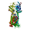
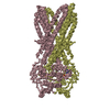
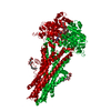
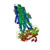
| Title | Cryo-EM structure of Patched in lipid nanodisc - the wildtype, 3.5 angstrom (re-processed with dataset of 7dzq) | ||||||||||||||||||||||||||||||
 Components Components | Protein patched homolog 1,Protein patched homolog 1 | ||||||||||||||||||||||||||||||
 Keywords Keywords | MEMBRANE PROTEIN / Caveolae / Hedgehog signaling / Lipid nanodisc / Patched / Ptc1 dimer | ||||||||||||||||||||||||||||||
| Function / homology |  Function and homology information Function and homology informationLigand-receptor interactions / Activation of SMO / neural plate axis specification / response to chlorate / cell differentiation involved in kidney development / hedgehog receptor activity / cell proliferation involved in metanephros development / neural tube patterning / smoothened binding / neural tube formation ...Ligand-receptor interactions / Activation of SMO / neural plate axis specification / response to chlorate / cell differentiation involved in kidney development / hedgehog receptor activity / cell proliferation involved in metanephros development / neural tube patterning / smoothened binding / neural tube formation / hedgehog family protein binding / hindlimb morphogenesis / epidermal cell fate specification / Hedgehog 'on' state / spinal cord motor neuron differentiation / prostate gland development / patched binding / negative regulation of cell division / somite development / Hedgehog 'off' state / limb morphogenesis / smooth muscle tissue development / pharyngeal system development / mammary gland duct morphogenesis / mammary gland epithelial cell differentiation / cellular response to cholesterol / pattern specification process / cell fate determination / metanephric collecting duct development / commissural neuron axon guidance / mammary gland development / response to alkaloid / dorsal/ventral pattern formation / regulation of growth / embryonic limb morphogenesis / negative regulation of multicellular organism growth / branching involved in ureteric bud morphogenesis / cholesterol binding / dorsal/ventral neural tube patterning / positive regulation of epidermal cell differentiation / dendritic growth cone / keratinocyte proliferation / epidermis development / positive regulation of cholesterol efflux / spermatid development / response to retinoic acid / embryonic organ development / negative regulation of osteoblast differentiation / response to mechanical stimulus / axonal growth cone / heart morphogenesis / regulation of mitotic cell cycle / liver regeneration / cyclin binding / animal organ morphogenesis / protein localization to plasma membrane / negative regulation of smoothened signaling pathway / neural tube closure / protein processing / brain development / caveola / negative regulation of epithelial cell proliferation / apical part of cell / response to estradiol / regulation of cell population proliferation / heparin binding / regulation of protein localization / glucose homeostasis / midbody / in utero embryonic development / postsynaptic membrane / cilium / response to xenobiotic stimulus / negative regulation of cell population proliferation / negative regulation of DNA-templated transcription / positive regulation of DNA-templated transcription / protein-containing complex binding / perinuclear region of cytoplasm / negative regulation of transcription by RNA polymerase II / Golgi apparatus / signal transduction / extracellular region / zinc ion binding / plasma membrane Similarity search - Function | ||||||||||||||||||||||||||||||
| Biological species |  | ||||||||||||||||||||||||||||||
| Method | ELECTRON MICROSCOPY / single particle reconstruction / cryo EM / Resolution: 3.5 Å | ||||||||||||||||||||||||||||||
 Authors Authors | Luo, Y. / Zhao, Y. / Qu, Q. / Li, D. | ||||||||||||||||||||||||||||||
| Funding support |  China, 4items China, 4items
| ||||||||||||||||||||||||||||||
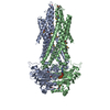
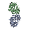
 Citation Citation | Journal: Structure / Year: 2021 Title: Cryo-EM study of patched in lipid nanodisc suggests a structural basis for its clustering in caveolae. Authors: Yitian Luo / Guoyue Wan / Xiang Zhang / Xuan Zhou / Qiuwen Wang / Jialin Fan / Hongmin Cai / Liya Ma / Hailong Wu / Qianhui Qu / Yao Cong / Yun Zhao / Dianfan Li / Abstract: The 12-transmembrane protein Patched (Ptc1) acts as a suppressor for Hedgehog (Hh) signaling by depleting sterols in the cytoplasmic membrane leaflet that are required for the activation of ...The 12-transmembrane protein Patched (Ptc1) acts as a suppressor for Hedgehog (Hh) signaling by depleting sterols in the cytoplasmic membrane leaflet that are required for the activation of downstream regulators. The positive modulator Hh inhibits Ptc1's transporter function by binding to Ptc1 and its co-receptors, which are locally concentrated in invaginated microdomains known as caveolae. Here, we reconstitute the mouse Ptc1 into lipid nanodiscs and determine its structure using single-particle cryoelectron microscopy. The structure is overall similar to those in amphipol and detergents but displays various conformational differences in the transmembrane region. Although most particles show monomers, we observe Ptc1 dimers with distinct interaction patterns and different membrane curvatures, some of which are reminiscent of caveolae. We find that an extramembranous "hand-shake" region rich in hydrophobic and aromatic residues mediates inter-Ptc1 interactions under different membrane curvatures. Our data provide a plausible framework for Ptc1 clustering in the highly curved caveolae. | ||||||||||||||||||||||||||||||
| History |
|
- Structure visualization
| Movie |
Movie viewer |
|---|---|
| Structure viewer | Molecule: MolmilJmol/JSmol |
- Downloads & links
-Download
| PDBx/mmCIF format | 7v6y.cif.gz | 190.4 KB | Display | PDBx/mmCIF format |
|---|---|---|---|---|
| PDB format | pdb7v6y.ent.gz | 140.9 KB | Display | PDB format |
| PDBx/mmJSON format | 7v6y.json.gz | Tree view | PDBx/mmJSON format | |
| Others |  Other downloads Other downloads |
-Validation report
| Arichive directory | https://data.pdbj.org/pub/pdb/validation_reports/v6/7v6yftp://data.pdbj.org/pub/pdb/validation_reports/v6/7v6y | HTTPS FTP |
|---|
-Related structure data
| Related structure data |  31753MC 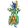 7v6zC M: map data used to model this data C: citing same article ( |
|---|---|
| Similar structure data |


-Links
 PDBj
PDBj


- Assembly
| Deposited unit | 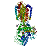
|
|---|---|
| 1 |
|
-Components
| #1: Protein | Mass: 121637.617 Da / Num. of mol.: 1 Source method: isolated from a genetically manipulated source Source: (gene. exp.)  Homo sapiens (human) / References: UniProt: Q61115 Homo sapiens (human) / References: UniProt: Q61115 | ||||||||
|---|---|---|---|---|---|---|---|---|---|
| #2: Polysaccharide | 2-acetamido-2-deoxy-beta-D-glucopyranose-(1-4)-2-acetamido-2-deoxy-beta-D-glucopyranose Source method: isolated from a genetically manipulated source | ||||||||
| #3: Chemical |   Mass: 386.654 Da / Num. of mol.: 2 / Source method: obtained synthetically / Formula: C27H46O Mass: 386.654 Da / Num. of mol.: 2 / Source method: obtained synthetically / Formula: C27H46O#4: Chemical | ChemComp-5VI / ( | 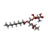  Mass: 481.474 Da / Num. of mol.: 1 / Source method: obtained synthetically / Formula: C20H36NO10P / Feature type: SUBJECT OF INVESTIGATION Mass: 481.474 Da / Num. of mol.: 1 / Source method: obtained synthetically / Formula: C20H36NO10P / Feature type: SUBJECT OF INVESTIGATION#5: Sugar |   Type: D-saccharide, beta linking / Mass: 221.208 Da / Num. of mol.: 3 / Source method: obtained synthetically / Formula: C8H15NO6 / Feature type: SUBJECT OF INVESTIGATION Type: D-saccharide, beta linking / Mass: 221.208 Da / Num. of mol.: 3 / Source method: obtained synthetically / Formula: C8H15NO6 / Feature type: SUBJECT OF INVESTIGATIONHas ligand of interest | Y | Has protein modification | Y | |
-Experimental details
-Experiment
| Experiment | Method: ELECTRON MICROSCOPY |
|---|---|
| EM experiment | Aggregation state: PARTICLE / 3D reconstruction method: single particle reconstruction |
- Sample preparation
| Component | Name: Ptc1 in lipid nanodisc / Type: COMPLEX / Entity ID: #1 / Source: RECOMBINANT | ||||||||||||||||||||||||||||||
|---|---|---|---|---|---|---|---|---|---|---|---|---|---|---|---|---|---|---|---|---|---|---|---|---|---|---|---|---|---|---|---|
| Molecular weight | Value: 160 kDa/nm / Experimental value: YES | ||||||||||||||||||||||||||||||
| Source (natural) | Organism: | ||||||||||||||||||||||||||||||
| Source (recombinant) | Organism: Homo sapiens (human) | ||||||||||||||||||||||||||||||
| Buffer solution | pH: 7.5 | ||||||||||||||||||||||||||||||
| Buffer component |
| ||||||||||||||||||||||||||||||
| Specimen | Conc.: 8.5 mg/ml / Embedding applied: NO / Shadowing applied: NO / Staining applied: NO / Vitrification applied: YES | ||||||||||||||||||||||||||||||
| Vitrification | Instrument: FEI VITROBOT MARK IV / Cryogen name: ETHANE / Humidity: 100 % / Chamber temperature: 277 K |
- Electron microscopy imaging
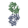
| Experimental equipment |  Model: Titan Krios / Image courtesy: FEI Company |
|---|---|
| Microscopy | Model: FEI TITAN KRIOS |
| Electron gun | Electron source:  FIELD EMISSION GUN / Accelerating voltage: 300 kV / Illumination mode: FLOOD BEAM FIELD EMISSION GUN / Accelerating voltage: 300 kV / Illumination mode: FLOOD BEAM |
| Electron lens | Mode: BRIGHT FIELD / Nominal magnification: 120000 X / Nominal defocus max: 2200 nm / Nominal defocus min: 1500 nm / Calibrated defocus min: 1100 nm / Calibrated defocus max: 3000 nm / Cs: 2.7 mm / C2 aperture diameter: 70 µm |
| Specimen holder | Cryogen: NITROGEN / Specimen holder model: FEI TITAN KRIOS AUTOGRID HOLDER |
| Image recording | Average exposure time: 2.23 sec. / Electron dose: 60 e/Å2 / Film or detector model: GATAN K3 (6k x 4k) / Num. of grids imaged: 1 |
| Image scans | Width: 5760 / Height: 4092 |
- Processing
| Software | Name: PHENIX / Version: 1.18.2_3874: / Classification: refinement | ||||||||||||||||||||||||||||||||||||||||
|---|---|---|---|---|---|---|---|---|---|---|---|---|---|---|---|---|---|---|---|---|---|---|---|---|---|---|---|---|---|---|---|---|---|---|---|---|---|---|---|---|---|
| EM software |
| ||||||||||||||||||||||||||||||||||||||||
| CTF correction | Type: NONE | ||||||||||||||||||||||||||||||||||||||||
| Particle selection | Num. of particles selected: 1363356 | ||||||||||||||||||||||||||||||||||||||||
| 3D reconstruction | Resolution: 3.5 Å / Resolution method: FSC 0.143 CUT-OFF / Num. of particles: 172299 / Algorithm: BACK PROJECTION / Symmetry type: POINT | ||||||||||||||||||||||||||||||||||||||||
| Atomic model building | B value: 57.04 / Protocol: FLEXIBLE FIT / Space: REAL | ||||||||||||||||||||||||||||||||||||||||
| Atomic model building | PDB-ID: 6MG8 Accession code: 6MG8 / Source name: PDB / Type: experimental model | ||||||||||||||||||||||||||||||||||||||||
| Refine LS restraints |
|

