 Movie
Movie Controller

[English] 日本語
 Yorodumi
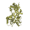
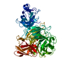
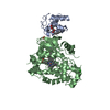
- PDB-7v6i: Crystal structure of lacto-N-biosidase BsaX from Bifidobacterium ... -
+ Open data

- Basic information
| Entry | Database: PDB / ID: 7v6i | |||||||||
|---|---|---|---|---|---|---|---|---|---|---|
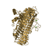
| Title | Crystal structure of lacto-N-biosidase BsaX from Bifidobacterium saguini, lacto-N-biose complex | |||||||||
 Components Components | Lacto-N-biosidase | |||||||||
 Keywords Keywords | HYDROLASE / Glycoside hydrolase / lacto-N-biosidase / lacto-N-biose complex | |||||||||
| Function / homology |  Function and homology information Function and homology information | |||||||||
| Biological species |  Bifidobacterium saguini DSM 23967 (bacteria) Bifidobacterium saguini DSM 23967 (bacteria) | |||||||||
| Method |  X-RAY DIFFRACTION / SYNCHROTRON / MOLECULAR REPLACEMENT / Resolution: 2.51 Å X-RAY DIFFRACTION / SYNCHROTRON / MOLECULAR REPLACEMENT / Resolution: 2.51 Å | |||||||||
 Authors Authors | Yamada, C. / Fushinobu, S. | |||||||||
| Funding support |  Japan, 2items Japan, 2items
| |||||||||
 Citation Citation | Journal: Biosci.Biotechnol.Biochem. / Year: 2022 Title: Crystal structures of glycoside hydrolase family 136 lacto-N-biosidases from monkey gut- and human adult gut bacteria. Authors: Yamada, C. / Katayama, T. / Fushinobu, S. | |||||||||
| History |
|
- Structure visualization
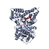
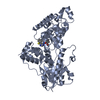
| Structure viewer | Molecule: MolmilJmol/JSmol |
|---|
- Downloads & links
-Download
| PDBx/mmCIF format | 7v6i.cif.gz | 133.1 KB | Display | PDBx/mmCIF format |
|---|---|---|---|---|
| PDB format | pdb7v6i.ent.gz | 98.7 KB | Display | PDB format |
| PDBx/mmJSON format | 7v6i.json.gz | Tree view | PDBx/mmJSON format | |
| Others |  Other downloads Other downloads |
-Validation report
| Arichive directory | https://data.pdbj.org/pub/pdb/validation_reports/v6/7v6iftp://data.pdbj.org/pub/pdb/validation_reports/v6/7v6i | HTTPS FTP |
|---|
-Related structure data
| Related structure data |  7v6mC  5gqfS S: Starting model for refinement C: citing same article ( |
|---|---|
| Similar structure data |


-Links
 PDBj
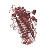
PDBj- Assembly
| Deposited unit | 
| ||||||||
|---|---|---|---|---|---|---|---|---|---|
| 1 |
| ||||||||
| Unit cell |
|
-Components
| #1: Protein | Mass: 68053.273 Da / Num. of mol.: 1 Source method: isolated from a genetically manipulated source Source: (gene. exp.) Bifidobacterium saguini DSM 23967 (bacteria)Gene: BISA_2023 / Plasmid: pET23b / Production host: |
|---|---|
| #2: Polysaccharide | beta-D-galactopyranose-(1-3)-2-acetamido-2-deoxy-beta-D-glucopyranose Source method: isolated from a genetically manipulated source |
| #3: Water | ChemComp-HOH /  Mass: 18.015 Da / Num. of mol.: 104 / Source method: isolated from a natural source / Formula: H2O Mass: 18.015 Da / Num. of mol.: 104 / Source method: isolated from a natural source / Formula: H2O |
| Has ligand of interest | N |
-Experimental details
-Experiment
| Experiment | Method: X-RAY DIFFRACTION / Number of used crystals: 1 |
|---|
- Sample preparation
| Crystal | Density Matthews: 2.24 Å3/Da / Density % sol: 45.02 % |
|---|---|
| Crystal grow | Temperature: 293 K / Method: vapor diffusion, sitting drop / pH: 4.6 Details: 0.5M ammonium sulfate, 0.1M sodium acetate (pH 4.6), 30% (w/v) PEG 4000 |
-Data collection
| Diffraction | Mean temperature: 100 K / Serial crystal experiment: N |
|---|---|
| Diffraction source | Source: SYNCHROTRON / Site: Photon Factory / Beamline: BL-5A / Wavelength: 1 Å |
| Detector | Type: DECTRIS PILATUS3 S 6M / Detector: PIXEL / Date: May 22, 2018 |
| Radiation | Monochromator: Numerical link type Si(111) double crystal monochromator Protocol: SINGLE WAVELENGTH / Monochromatic (M) / Laue (L): M / Scattering type: x-ray |
| Radiation wavelength | Wavelength: 1 Å / Relative weight: 1 |
| Reflection | Resolution: 2.51→49.08 Å / Num. obs: 20374 / % possible obs: 100 % / Redundancy: 10 % / Biso Wilson estimate: 15.86 Å2 / CC1/2: 0.995 / Rmerge(I) obs: 0.184 / Rpim(I) all: 0.061 / Rrim(I) all: 0.194 / Net I/σ(I): 11.2 |
| Reflection shell | Resolution: 2.51→2.61 Å / Redundancy: 10.2 % / Rmerge(I) obs: 0.695 / Mean I/σ(I) obs: 3.8 / Num. unique obs: 2262 / CC1/2: 0.913 / Rpim(I) all: 0.228 / Rrim(I) all: 0.732 / % possible all: 100 |
- Processing
| Software |
| ||||||||||||||||||||||||||||||||||||||||||||||||||||||||||||||||||||||||||||||||||||||||||||||||||||||||||||||||||||||||||||||||||||||||||||||||||||||||||||||||||||||||||||||||||||||
|---|---|---|---|---|---|---|---|---|---|---|---|---|---|---|---|---|---|---|---|---|---|---|---|---|---|---|---|---|---|---|---|---|---|---|---|---|---|---|---|---|---|---|---|---|---|---|---|---|---|---|---|---|---|---|---|---|---|---|---|---|---|---|---|---|---|---|---|---|---|---|---|---|---|---|---|---|---|---|---|---|---|---|---|---|---|---|---|---|---|---|---|---|---|---|---|---|---|---|---|---|---|---|---|---|---|---|---|---|---|---|---|---|---|---|---|---|---|---|---|---|---|---|---|---|---|---|---|---|---|---|---|---|---|---|---|---|---|---|---|---|---|---|---|---|---|---|---|---|---|---|---|---|---|---|---|---|---|---|---|---|---|---|---|---|---|---|---|---|---|---|---|---|---|---|---|---|---|---|---|---|---|---|---|
| Refinement | Method to determine structure: MOLECULAR REPLACEMENT Starting model: 5GQF Resolution: 2.51→49.08 Å / Cor.coef. Fo:Fc: 0.958 / Cor.coef. Fo:Fc free: 0.897 / SU B: 8.99 / SU ML: 0.199 / Cross valid method: THROUGHOUT / ESU R: 1.391 / ESU R Free: 0.287 / Stereochemistry target values: MAXIMUM LIKELIHOOD / Details: HYDROGENS HAVE BEEN ADDED IN THE RIDING POSITIONS
| ||||||||||||||||||||||||||||||||||||||||||||||||||||||||||||||||||||||||||||||||||||||||||||||||||||||||||||||||||||||||||||||||||||||||||||||||||||||||||||||||||||||||||||||||||||||
| Solvent computation | Ion probe radii: 0.8 Å / Shrinkage radii: 0.8 Å / VDW probe radii: 1.2 Å / Solvent model: MASK | ||||||||||||||||||||||||||||||||||||||||||||||||||||||||||||||||||||||||||||||||||||||||||||||||||||||||||||||||||||||||||||||||||||||||||||||||||||||||||||||||||||||||||||||||||||||
| Displacement parameters | Biso mean: 25.598 Å2
| ||||||||||||||||||||||||||||||||||||||||||||||||||||||||||||||||||||||||||||||||||||||||||||||||||||||||||||||||||||||||||||||||||||||||||||||||||||||||||||||||||||||||||||||||||||||
| Refinement step | Cycle: 1 / Resolution: 2.51→49.08 Å
| ||||||||||||||||||||||||||||||||||||||||||||||||||||||||||||||||||||||||||||||||||||||||||||||||||||||||||||||||||||||||||||||||||||||||||||||||||||||||||||||||||||||||||||||||||||||
| Refine LS restraints |
|